20
views
0
recommends
+1 Recommend
0 collections
    0
    shares
      • Record: found
      • Abstract: found
      • Article: found
      Is Open Access

      Effect of Intraocular Forward Scattering and Corneal Higher-Order Aberrations on Visual Acuity after Descemet’s Stripping Automated Endothelial Keratoplasty

      research-article

      Read this article at

      Bookmark
          There is no author summary for this article yet. Authors can add summaries to their articles on ScienceOpen to make them more accessible to a non-specialist audience.

          Abstract

          Purpose

          To assess the relationship of intraocular forward scattering and corneal higher-order aberrations (HOAs) with best spectacle corrected visual acuity (BSCVA) after Descemet’s stripping automated endothelial keratoplasty (DSAEK), and to compare these parameters between DSAEK and non-Descemet’s stripping automated endothelial keratoplasty (n-DSAEK) groups.

          Methods

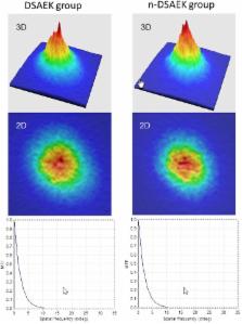
          This retrospective study enrolled thirty eyes of 30 consecutive patients who underwent standard DSAEK, and who underwent successful phacoemulsification with intraocular lens implantation before DSAEK. The mean age at the time of surgery was 71.7 ± 10.4 years. We quantitatively evaluated the objective scattering index (OSI) using the double-pass instrument (OQAS II, Visiometrics) and corneal HOAs using Hartmann-Shack aberrometry (KR-9000PW, Topcon) 3 months postoperatively.

          Results

          The mean OSI, corneal HOAs, and logMAR BSCVA 3 months after DSAEK were 7.91 ± 3.58, 0.43 ± 0.27 μm, and 0.32 ± 0.25, respectively. We found a significant correlation between the OSI and logMAR BSCVA (Spearman correlation coefficient r=0.714, p<0.001), but no significant association between corneal HOAs and logMAR BSCVA 3 months postoperatively (r=0.209, p=0.267). We found no significant differences in any postoperative parameters between the DSAEK and n-DSAEK groups (p>0.05).

          Conclusions

          Our pilot study demonstrated that the postoperative corrected visual acuity was significantly correlated with intraocular forward scattering, but not with corneal HOAs in post-DSAEK eyes, suggesting that intraocular forward scattering plays a more essential role in postoperative visual performance than corneal aberrations after DSAEK. The detailed visual performance, such as HOAs and intraocular scattering, after n-DSAEK appears to be essentially equivalent to that after DSAEK.

          Related collections

          Most cited references20

          • Record: found
          • Abstract: found
          • Article: not found

          Descemet-stripping automated endothelial keratoplasty.

          To evaluate the speed of visual recovery in 16 consecutive patients with corneal endothelial dysfunction who received Descemet-stripping automated endothelial keratoplasty (DSAEK). This is a retrospective study of a novel method for small-incision endothelial transplantation (DSAEK). Endothelial replacement was accomplished with Descemet stripping of the recipient and insertion of a posterior donor tissue that had been prepared with a microkeratome. Best spectacle-corrected visual acuity (BSCVA) by manifest refraction, endothelial counts, and dislocation rates were measured up to 12 months after DSAEK. Sixteen consecutive patients underwent uncomplicated DSAEK. Three patients had known optic nerve or macular disease precluding vision better than 20/200. Of the remaining 14 patients, 11 had BSCVA of 20/40 by postoperative week 12 (7 by week 6). The remaining 2 were 20/50 by weeks 6 and 12. All 14 patients were 20/40 or better at 1 year. One patient had a primary graft failure, and surgery was repeated with 20/40 BSCVA at 1 year. The dislocation rate was 25%. The average cell count between 7 and 10 months was 1714. The average pachymetry was 682. DSAEK surgery allows rapid, excellent BSCVA visual recovery. The rate of visual recovery is more rapid than usually found with penetrating keratoplasty.
            Bookmark
            • Record: found
            • Abstract: found
            • Article: found
            Is Open Access

            An Objective Scatter Index Based on Double-Pass Retinal Images of a Point Source to Classify Cataracts

            Purpose To propose a new objective scatter index (OSI) based in the analysis of double-pass images of a point source to rank and classify cataract patients. This classification scheme is compared with a current subjective system. Methods We selected a population including a group of normal young eyes as control and patients diagnosed with cataract (grades NO2, NO3 and NO4) according to the Lens Opacities Classification System (LOCS III). For each eye, we recorded double-pass retinal images of a point source. In each patient, we determined an objective scatter index (OSI) as the ratio of the intensity at an eccentric location in the image and the central part. This index provides information on the relevant forward scatter affecting vision. Since the double-pass retinal images are affected by both ocular aberrations and intraocular scattering, an analysis was performed to show the ranges of contributions of aberrations to the OSI. Results We used the OSI values to classify each eye according to the degree of scatter. The young normal eyes of the control group had OSI values below 1, while the OSI for subjects in LOCS grade II were around 1 to 2. The use of the objective index showed some of the weakness of subjective classification schemes. In particular, several subjects initially classified independently as grade NO2 or NO3 had similar OSI values, and in some cases even higher than subjects classified as grade NO4. A new classification scheme based in OSI is proposed. Conclusions We introduced an objective index based in the analysis of double-pass retinal images to classify cataract patients. The method is robust and fully based in objective measurements; i.e., not depending on subjective decisions. This procedure could be used in combination with standard current methods to improve cataract patient surgery scheduling.
              Bookmark
              • Record: found
              • Abstract: found
              • Article: not found

              Descemet's stripping with endothelial keratoplasty in 50 eyes: a refractive neutral corneal transplant.

              To evaluate early visual and refractive outcomes following treatment of corneal endothelial dysfunction with a corneal transplantation technique, Descemet's stripping with endothelial keratoplasty (DSEK). Visual and refractive outcomes of the first 50 consecutive cases of DSEK performed by a single surgeon between December 2003 and July 2004 were analyzed retrospectively. The DSEK technique consisted of stripping Descemet's membrane and endothelium from a recipient cornea and transplanting the posterior stroma and endothelium of a donor cornea through a 5-mm incision. Results are reported for 50 eyes in 47 patients (30 women and 17 men). Mean patient age at surgery was 70 +/- 12 years (range: 34 to 89 years). Five eyes were treated for corneal edema or bullous keratopathy and 45 for Fuchs' endothelial dystrophy. Seven eyes were phakic and 43 were pseudophakic. Six months after surgery, mean manifest cylinder was 1.5 +/- 0.94 diopters (D), unchanged from preoperative cylinder of 1.5 +/- 1.0 D. Mean manifest spherical equivalent refraction was 0.15 +/- 1.5 D at 6-month follow-up compared with -0.36 +/- 1.4 D preoperatively (P = .10) At 3- and 6-month follow-up, significant improvement was noted in mean best spectacle-corrected visual acuity compared with the preoperative mean of 20/100 (P = .007). At 6-month follow-up, 31 (62%) eyes refracted to > or = 20/40 and 38 (76%) eyes saw > or = 20/50. Compared to standard penetrating keratoplasty, DSEK causes minimal refractive change and provides rapid visual recovery for patients with endothelial dysfunction. This technique maintains the structural integrity of the cornea by preserving the recipient's epithelium, Bowman's layer, and entire stromal thickness.
                Bookmark

                Author and article information

                Contributors
                Role: Editor
                Journal
                PLoS One
                PLoS ONE
                plos
                plosone
                PLoS ONE
                Public Library of Science (San Francisco, CA USA )
                1932-6203
                19 June 2015
                2015
                : 10
                : 6
                : e0131110
                Affiliations
                [001]Department of Ophthalmology, University of Kitasato School of Medicine, Kanagawa, Japan
                Sun Yat-sen University, CHINA
                Author notes

                Competing Interests: The authors have declared that no competing interests exist.

                Conceived and designed the experiments: KK KS HK AI. Performed the experiments: KK HA KS HK AI. Analyzed the data: KK HA HK AI. Contributed reagents/materials/analysis tools: KK KS. Wrote the paper: KK.

                Article
                PONE-D-14-50037
                10.1371/journal.pone.0131110
                4474429
                26090889
                3a682737-613a-4275-93d2-04e0b05535d4
                Copyright @ 2015

                This is an open access article distributed under the terms of the Creative Commons Attribution License, which permits unrestricted use, distribution, and reproduction in any medium, provided the original author and source are credited

                History
                : 6 November 2014
                : 28 May 2015
                Page count
                Figures: 3, Tables: 2, Pages: 10
                Funding
                The authors received no specific funding for this work.
                Categories
                Research Article
                Custom metadata
                The authors confirm that all data underlying the findings are fully available without restriction. All relevant data are within the paper.

                Uncategorized
                Uncategorized

                Comments

                Comment on this article