34
views
0
recommends
+1 Recommend
0 collections
    0
    shares
      • Record: found
      • Abstract: found
      • Article: found
      Is Open Access

      An Objective Scatter Index Based on Double-Pass Retinal Images of a Point Source to Classify Cataracts

      research-article

      Read this article at

      Bookmark
          There is no author summary for this article yet. Authors can add summaries to their articles on ScienceOpen to make them more accessible to a non-specialist audience.

          Abstract

          Purpose

          To propose a new objective scatter index (OSI) based in the analysis of double-pass images of a point source to rank and classify cataract patients. This classification scheme is compared with a current subjective system.

          Methods

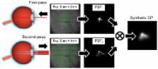
          We selected a population including a group of normal young eyes as control and patients diagnosed with cataract (grades NO2, NO3 and NO4) according to the Lens Opacities Classification System (LOCS III). For each eye, we recorded double-pass retinal images of a point source. In each patient, we determined an objective scatter index (OSI) as the ratio of the intensity at an eccentric location in the image and the central part. This index provides information on the relevant forward scatter affecting vision. Since the double-pass retinal images are affected by both ocular aberrations and intraocular scattering, an analysis was performed to show the ranges of contributions of aberrations to the OSI.

          Results

          We used the OSI values to classify each eye according to the degree of scatter. The young normal eyes of the control group had OSI values below 1, while the OSI for subjects in LOCS grade II were around 1 to 2. The use of the objective index showed some of the weakness of subjective classification schemes. In particular, several subjects initially classified independently as grade NO2 or NO3 had similar OSI values, and in some cases even higher than subjects classified as grade NO4. A new classification scheme based in OSI is proposed.

          Conclusions

          We introduced an objective index based in the analysis of double-pass retinal images to classify cataract patients. The method is robust and fully based in objective measurements; i.e., not depending on subjective decisions. This procedure could be used in combination with standard current methods to improve cataract patient surgery scheduling.

          Related collections

          Most cited references16

          • Record: found
          • Abstract: found
          • Article: not found

          Straylight effects with aging and lens extraction.

          To assess possible gains and losses in straylight values among the population to consider straylight as added benefit of lens extraction. In this cross-sectional design, data from a multicenter study on visual function in automobile drivers were analyzed. On both eyes of 2,422 subjects, visual acuity (logarithm of the minimum angle of resolution [logMAR] in steps of 0.02 log units), straylight on the retina (psychophysical compensation comparison method), and lens opacity (slit-lamp scoring using the Lens Opacities Classification System III [LOCS III] system) were determined. Three groups were defined: 220 pseudophakic eyes, 3,182 noncataractous eyes (average LOCS III score, 3.0). Noncataractous straylight values increases strongly with age as: log(s) = constant + log(1 + (age / 65)(4)), doubling by the age of 65 years, and tripling by the age of 77 years. Population standard deviation around this age norm was approximately 0.10 log units. The cataract eyes (in this active driver group) had relatively mild straylight increase. In pseudophakia, straylight values may be very good, better even than in the noncataract group. Visual acuity and straylight were found to vary quite independently. Lens extraction holds promise not only to improve on the condition of the cataract eye, but also to improve on the age-normal eye. Lens extraction potentially reverses the strong age increase in straylight value, quite independently from visual acuity.
            Bookmark
            • Record: found
            • Abstract: found
            • Article: not found

            Compensation comparison method for assessment of retinal straylight.

            Presently, no instrument or method exists that is generally accepted for routine clinical assessment of (functional) retinal straylight. Yet retinal straylight is the cause of major patient complaints, such as hindrance from glare and loss of contrast. It results from disturbances in the optical media that increase light-scattering over angles of 1 degrees to 90 degrees . Its assessment would help to decide whether to perform surgery for (early) cataract and would help in the evaluation of corneal or vitreal turbidity. The psychophysical technique of the "direct compensation" method was adapted to make it suitable for routine clinical assessment. In the new approach, called "compensation comparison, " the central test field is subdivided into two half fields: one with and one without counterphase compensation light. The subject's task is a forced-choice comparison between the two half fields, to decide which half flickers more strongly. A theoretical form for the respective psychometric function was defined and experimentally verified in a laboratory experiment involving seven subjects, with and without artificially increased light scattering. The method was applied in a separate multicenter study. Its reliability was additionally tested with a commercial implement (C-Quant; Oculus Optikgeräte, Wetzlar-Dutenhofen, Germany). A repeated-measures SD of 0.07 log units was achieved, to be compared with differences in the young normal population of 0.4 log units and an increase with healthy aging by 0.5 log units at 80 years and by 1.0 or more log units with (early) cataract or corneal disturbances. Reliability was further found to be high when using the commercial version of the The compensation comparison method for measuring retinal straylight is suited for clinical use to diagnose patients with complaints caused by large angle light scattering in the eye such as early cataract.
              Bookmark
              • Record: found
              • Abstract: found
              • Article: not found

              Effects of aging in retinal image quality.

              The retinal image quality characterized by the modulation-transfer function of the eye was measured for two groups of subjects aged in the late twenties and mid sixties, respectively. In both groups, we obtained modulation transfer functions by using a double-pass method under the same experimental conditions: 4-mm artificial pupil, paralyzed accommodation, and objective control of the refractive state and centering. Results showed lower values of modulation in the retinal image for older subjects compared with the younger subjects. The modulation transfer function ratio is similar to that previously found for contrast-sensitivity measurements with subjects in the same age groups. These results suggest that a significant fraction of the loss in spatial vision with age has an optical origin. Apart from the well-known increase in intraocular scattering, there also appears to be an increment in ocular aberration that causes an additional reduction in the contrast of retinal images.
                Bookmark

                Author and article information

                Contributors
                Role: Editor
                Journal
                PLoS One
                plos
                plosone
                PLoS ONE
                Public Library of Science (San Francisco, USA )
                1932-6203
                2011
                4 February 2011
                : 6
                : 2
                : e16823
                Affiliations
                [1 ]Laboratorio de Óptica, Universidad de Murcia, Murcia, Spain
                [2 ]Hospital Universitario Virgen de la Arrixaca, Murcia, Spain
                [3 ]Centre de Desenvolupament de Sensors, Instrumentació i Sistemes (CD6), Universidad Politécnica de Cataluña, Tarrasa, Barcelona, Spain
                The University of Hong Kong, Hong Kong
                Author notes

                Conceived and designed the experiments: PA. Performed the experiments: AB GP EA ADC. Analyzed the data: PA AB GP EA ADC JP JMM. Contributed reagents/materials/analysis tools: JP JMM. Wrote the paper: PA.

                Article
                PONE-D-10-03387
                10.1371/journal.pone.0016823
                3033912
                21326868
                ca70f17b-873d-41a0-8d06-0019593da656
                Artal et al. This is an open-access article distributed under the terms of the Creative Commons Attribution License, which permits unrestricted use, distribution, and reproduction in any medium, provided the original author and source are credited.
                History
                : 11 October 2010
                : 11 January 2011
                Page count
                Pages: 7
                Categories
                Research Article
                Biology
                Anatomy and Physiology
                Ocular System
                Ocular Anatomy
                Engineering
                Bioengineering
                Medical Devices
                Materials Science
                Material Properties
                Optical Properties
                Medicine
                Anatomy and Physiology
                Ocular System
                Ocular Anatomy
                Diagnostic Medicine
                Test Evaluation
                Ophthalmology
                Cataracts and Other Lens Disorders
                Surgery
                Ophthalmology
                Physics
                Condensed-Matter Physics
                Optics

                Uncategorized
                Uncategorized

                Comments

                Comment on this article