18
views
0
recommends
+1 Recommend
0 collections
    0
    shares
      • Record: found
      • Abstract: found
      • Article: found
      Is Open Access

      Effect of Light Scattering and Higher-order Aberrations on Visual Performance in Eyes with Granular Corneal Dystrophy

      research-article

      Read this article at

      Bookmark
          There is no author summary for this article yet. Authors can add summaries to their articles on ScienceOpen to make them more accessible to a non-specialist audience.

          Abstract

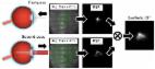
          This study was aimed to assess the relationship of intraocular forward scattering, corneal backward scattering, and corneal higher-order aberrations (HOAs) with corrected distance visual acuity (CDVA) in eyes with granular corneal dystrophy (GCD). We retrospectively examined forty two eyes of 42 consecutive patients who diagnosed GCD, and age-matched 20 eyes of 20 healthy subjects. We assessed objective scattering index (OSI) using the double-pass instrument (OQAS II, Visiometrics), corneal densitometry (CD) using the Scheimpflug rotating camera (Pentacam HR, Oculus), and corneal HOAs using the Hartmann-Shack aberrometry (KR-9000, Topcon). The OSI, CD, and corneal HOAs were significantly larger in the GCD group than those in the control group (Mann-Whitney U test, p < 0.001). We found significant correlations of logMAR CDVA with the OSI (Spearman correlation coefficient r = 0.577, p < 0.001), and with the CD (r = 0.340, p = 0.028), but no significant association with corneal HOAs (r = 0.061, p = 0.701). Intraocular forward scattering, corneal backward scattering, and corneal HOAs in eyes with GCD were higher than that in normal eyes. The CDVA was significantly correlated with intraocular forward scattering, but not with corneal HOAs in eyes with GCD, suggesting that light scattering, especially forward scattering, plays a more vital role in visual performance than corneal aberrations in eyes with GCD.

          Related collections

          Most cited references14

          • Record: found
          • Abstract: found
          • Article: found
          Is Open Access

          An Objective Scatter Index Based on Double-Pass Retinal Images of a Point Source to Classify Cataracts

          Purpose To propose a new objective scatter index (OSI) based in the analysis of double-pass images of a point source to rank and classify cataract patients. This classification scheme is compared with a current subjective system. Methods We selected a population including a group of normal young eyes as control and patients diagnosed with cataract (grades NO2, NO3 and NO4) according to the Lens Opacities Classification System (LOCS III). For each eye, we recorded double-pass retinal images of a point source. In each patient, we determined an objective scatter index (OSI) as the ratio of the intensity at an eccentric location in the image and the central part. This index provides information on the relevant forward scatter affecting vision. Since the double-pass retinal images are affected by both ocular aberrations and intraocular scattering, an analysis was performed to show the ranges of contributions of aberrations to the OSI. Results We used the OSI values to classify each eye according to the degree of scatter. The young normal eyes of the control group had OSI values below 1, while the OSI for subjects in LOCS grade II were around 1 to 2. The use of the objective index showed some of the weakness of subjective classification schemes. In particular, several subjects initially classified independently as grade NO2 or NO3 had similar OSI values, and in some cases even higher than subjects classified as grade NO4. A new classification scheme based in OSI is proposed. Conclusions We introduced an objective index based in the analysis of double-pass retinal images to classify cataract patients. The method is robust and fully based in objective measurements; i.e., not depending on subjective decisions. This procedure could be used in combination with standard current methods to improve cataract patient surgery scheduling.
            Bookmark
            • Record: found
            • Abstract: found
            • Article: not found

            Normative values for corneal densitometry analysis by Scheimpflug optical assessment.

            To describe the normative data for corneal Scheimpflug densitometry based on a cohort of normal participants. A total of 445 healthy participants were recruited for assessment (794 eyes). Left and right eyes were considered separately. All participants were assessed using the corneal densitometry analysis add-on to the standard software of the Oculus Pentacam. Densitometry measurements were obtained and expressed in standardized grayscale units (GSU). All participants were Caucasian; 42% were male and 58% were female. The mean age was 48.0 ± 15.3 years (range, 20.2-84.2 years). Mean corneal densitometry over the 12-mm-diameter area was 19.74 ± 3.89 GSU. When divided by radial zone, densitometry values were lowest in the central zone (16.76 ± 1.87 GSU) and highest in the periphery (27.36 ± 7.47 GSU). There was no difference between central zone and the surrounding 2- to 6-mm annulus (P > 0.05), though the 6- to 10-mm and the 10- to 12-mm zones displayed higher densitometry values (P < 0.001). When divided by depth, the anterior layer displayed the highest densitometry reading of 25.81 ± 5.14 GSU, which was significantly higher than that of both the central (P < 0.001) and the posterior layers (P < 0.001). Changes in corneal densitometry were correlated with age, though not within the central 6-mm-diameter ring. No sex difference was seen within the cohort. This add-on to the standard imaging software allows rapid and objective assessment of the corneal densitometry. We provide normative data that may be used as a reference facilitating research and complementing clinical examination.
              Bookmark
              • Record: found
              • Abstract: found
              • Article: not found

              Determination of treatment strategies for granular corneal dystrophy type 2 using Fourier-domain optical coherence tomography.

              To evaluate the use of Fourier-domain optical coherence tomography (FD-OCT) in the selection and planning of surgical procedures to treat GCD2 (granular corneal dystrophy type 2). An RTVue-100 FD-OCT (Optovue, Fremont, California) was used to determine the size, depth and location of deposits in six patients with homozygous or heterozygous GCD2. The RTVue-100 FD-OCT revealed the depth of the deposits quite precisely, allowing the determination of the appropriate depth of PTK (phototherapeutic keratectomy) and the appropriate selection of lamellar versus penetrating keratoplasty as the procedure of choice. In each case, visually significant opacities were adequately removed or would be removed. FD-OCT provides useful information for the selection and planning of surgical procedures to remove corneal opacities in patients with GCD2.
                Bookmark

                Author and article information

                Journal
                Sci Rep
                Sci Rep
                Scientific Reports
                Nature Publishing Group
                2045-2322
                18 April 2016
                2016
                : 6
                : 24677
                Affiliations
                [1 ]Department of Ophthalmology, University of Kitasato School of Medicine , Japan
                [2 ]Kitasato University School of Allied Health Sciences , Kanagawa, Japan
                Author notes
                Article
                srep24677
                10.1038/srep24677
                4834530
                27086550
                0a76b2b3-19fa-4ee9-845f-60e7c362d52f
                Copyright © 2016, Macmillan Publishers Limited

                This work is licensed under a Creative Commons Attribution 4.0 International License. The images or other third party material in this article are included in the article’s Creative Commons license, unless indicated otherwise in the credit line; if the material is not included under the Creative Commons license, users will need to obtain permission from the license holder to reproduce the material. To view a copy of this license, visit http://creativecommons.org/licenses/by/4.0/

                History
                : 14 January 2016
                : 04 April 2016
                Categories
                Article

                Uncategorized
                Uncategorized

                Comments

                Comment on this article