10
views
0
recommends
+1 Recommend
0 collections
    0
    shares
      • Record: found
      • Abstract: found
      • Article: found
      Is Open Access

      Characterization of developmental and molecular factors underlying release heterogeneity at Drosophila synapses

      research-article

      Read this article at

      Bookmark
          There is no author summary for this article yet. Authors can add summaries to their articles on ScienceOpen to make them more accessible to a non-specialist audience.

          Abstract

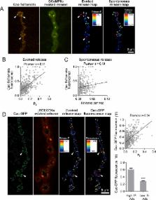
          Neurons communicate through neurotransmitter release at specialized synaptic regions known as active zones (AZs). Using biosensors to visualize single synaptic vesicle fusion events at Drosophila neuromuscular junctions, we analyzed the developmental and molecular determinants of release probability ( P r ) for a defined connection with ~300 AZs. P r was heterogeneous but represented a stable feature of each AZ. P r remained stable during high frequency stimulation and retained heterogeneity in mutants lacking the Ca 2+ sensor Synaptotagmin 1. P r correlated with both presynaptic Ca 2+ channel abundance and Ca 2+ influx at individual release sites. P r heterogeneity also correlated with glutamate receptor abundance, with high P r connections developing receptor subtype segregation. Intravital imaging throughout development revealed that AZs acquire high P r during a multi-day maturation period, with P r heterogeneity largely reflecting AZ age. The rate of synapse maturation was activity-dependent, as both increases and decreases in neuronal activity modulated glutamate receptor field size and segregation.

          eLife digest

          To send a message to its neighbor, a neuron releases chemicals called neurotransmitters into the gap – or synapse – between them. The neurotransmitter molecules bind to proteins on the receiver neuron called receptors. But what causes the sender neuron to release neurotransmitter in the first place? The process starts when an electrical impulse called an action potential arrives at the sender cell. Its arrival causes channels in the membrane of the sender neuron to open, so that calcium ions flood into the cell. The calcium ions interact with packages of neurotransmitter molecules, known as synaptic vesicles. This causes some of the vesicles to empty their contents into the synapse.

          But this process is not particularly reliable. Only a small fraction of action potentials cause vesicles to fuse with the synaptic membrane. How likely this is to occur varies greatly between neurons, and even between synapses formed by the same neuron. Synapses that are likely to release neurotransmitter are said to be strong. They are good at passing messages from the sender neuron to the receiver. Synapses with a low probability of release are said to be weak. But what exactly differs between strong and weak synapses?

          Akbergenova et al. studied synapses between motor neurons and muscle cells in the fruit fly Drosophila. Each motor neuron forms several hundred synapses. Some of these synapses are 50 times more likely to release neurotransmitter than others. Using calcium imaging and genetics, Akbergenova et al. showed that sender cells at strong synapses have more calcium channels than sender cells at weak synapses. The subtypes and arrangement of receptor proteins also differ between the receiver neurons of strong versus weak synapses. Finally, studies in larvae revealed that newly formed synapses all start out weak and then gradually become stronger. How fast this strengthening occurs depends on how active the neuron at the synapse is.

          This study has shown, in unprecedented detail, key molecular factors that make some fruit fly synapses more likely to release neurotransmitter than others. Many proteins at synapses of mammals resemble those at fruit fly synapses. This means that similar factors may also explain differences in synaptic strength in the mammalian brain. Changes in the strength of synapses underlie the ability to learn. Furthermore, many neurological and psychiatric disorders result from disruption of synapses. Understanding the molecular basis of synapses will thus provide clues to the origins of certain brain diseases.

          Related collections

          Most cited references111

          • Record: found
          • Abstract: found
          • Article: found
          Is Open Access

          Sensitive red protein calcium indicators for imaging neural activity

          Genetically encoded calcium indicators (GECIs) allow measurement of activity in large populations of neurons and in small neuronal compartments, over times of milliseconds to months. Although GFP-based GECIs are widely used for in vivo neurophysiology, GECIs with red-shifted excitation and emission spectra have advantages for in vivo imaging because of reduced scattering and absorption in tissue, and a consequent reduction in phototoxicity. However, current red GECIs are inferior to the state-of-the-art GFP-based GCaMP6 indicators for detecting and quantifying neural activity. Here we present improved red GECIs based on mRuby (jRCaMP1a, b) and mApple (jRGECO1a), with sensitivity comparable to GCaMP6. We characterized the performance of the new red GECIs in cultured neurons and in mouse, Drosophila, zebrafish and C. elegans in vivo. Red GECIs facilitate deep-tissue imaging, dual-color imaging together with GFP-based reporters, and the use of optogenetics in combination with calcium imaging. DOI: http://dx.doi.org/10.7554/eLife.12727.001
            Bookmark
            • Record: found
            • Abstract: found
            • Article: not found

            Quantitative ultrastructural analysis of hippocampal excitatory synapses.

            From three-dimensional reconstructions of CA1 excitatory synapses in the rodent hippocampus and in culture, we have estimated statistical distributions of active zone and postsynaptic density (PSD) sizes (average area approximately 0.04 micron2), the number of active zones per bouton (usually one), the number of docked vesicles per active zone (approximately 10), and the total number of vesicles per bouton (approximately 200), and we have determined relationships between these quantities, all of which vary from synapse to synapse but are highly correlated. These measurements have been related to synaptic physiology. In particular, we propose that the distribution of active zone areas can account for the distribution of synaptic release probabilities and that each active zone constitutes a release site as identified in the standard quantal theory attributable to Katz (1969).
              Bookmark
              • Record: found
              • Abstract: found
              • Article: not found

              Release probability of hippocampal glutamatergic terminals scales with the size of the active zone

              Cortical synapses display remarkable structural, molecular and functional heterogeneity. Our knowledge regarding the relationship between the ultrastructural and functional parameters is still fragmented. Here we asked how the release probability and presynaptic [Ca2+] transients relate to the ultrastructure of rat hippocampal glutamatergic axon terminals. Two-photon Ca2+ imaging-derived optical quantal analysis and correlated electron microscopic reconstructions revealed a tight correlation between the release probability and the active zone area. The peak amplitude of [Ca2+] transients in single boutons also positively correlated with the active zone area. Freeze-fracture immunogold labeling revealed that the voltage-gated Ca2+ channel subunit Cav2.1 and the presynaptic protein Rim1/2 are confined to the active zone and their numbers scale linearly with the active zone area. Gold particles for Cav2.1 showed a nonrandom distribution within the active zones. Our results demonstrate that the number of several active zone proteins, including presynaptic Ca2+ channels, docked vesicles and the release probability scales linearly with the active zone area.
                Bookmark

                Author and article information

                Contributors
                Role: Senior Editor
                Role: Reviewing Editor
                Journal
                eLife
                Elife
                eLife
                eLife
                eLife Sciences Publications, Ltd
                2050-084X
                10 July 2018
                2018
                : 7
                : e38268
                Affiliations
                [1 ]deptThe Picower Institute for Learning and Memory Massachusetts Institute of Technology CambridgeUnited States
                [2 ]deptDepartment of Biology Massachusetts Institute of Technology CambridgeUnited States
                [3 ]deptDepartment of Brain and Cognitive Sciences Massachusetts Institute of Technology CambridgeUnited States
                Brandeis University United States
                Baylor College of Medicine United States
                Baylor College of Medicine United States
                University of Miami Miller School of Medicine United States
                University of Southern California United States
                Author notes

                The authors declare no competing interests.

                Author information
                http://orcid.org/0000-0001-5576-2887
                Article
                38268
                10.7554/eLife.38268
                6075867
                29989549
                e8115c31-364e-497d-b0f6-4f6f034df37b
                © 2018, Akbergenova et al

                This article is distributed under the terms of the Creative Commons Attribution License, which permits unrestricted use and redistribution provided that the original author and source are credited.

                History
                : 11 May 2018
                : 30 June 2018
                Funding
                Funded by: FundRef http://dx.doi.org/10.13039/100000002, National Institutes of Health;
                Award ID: MH104536
                Award Recipient :
                Funded by: FundRef http://dx.doi.org/10.13039/100000002, National Institutes of Health;
                Award ID: T32GM007287
                Award Recipient :
                The funders had no role in study design, data collection and interpretation, or the decision to submit the work for publication.
                Categories
                Research Article
                Neuroscience
                Custom metadata
                Active zone release probability is correlated with calcium channel density and calcium influx at single release sites, with release strength increasing in an activity-dependent manner during synapse maturation.

                Life sciences
                active zone,synapse,calcium channels,synaptic plasticity,synapse development,glutamate receptors,d. melanogaster

                Comments

                Comment on this article