23
views
0
recommends
+1 Recommend
0 collections
    0
    shares
      • Record: found
      • Abstract: found
      • Article: found
      Is Open Access

      Repairing the brain with physical exercise: Cortical thickness and brain volume increases in long-term pediatric brain tumor survivors in response to a structured exercise intervention

      research-article

      Read this article at

      Bookmark
          There is no author summary for this article yet. Authors can add summaries to their articles on ScienceOpen to make them more accessible to a non-specialist audience.

          Abstract

          There is growing evidence that exercise induced experience dependent plasticity may foster structural and functional recovery following brain injury. We examined the efficacy of exercise training for neural and cognitive recovery in long-term pediatric brain tumor survivors treated with radiation.

          We conducted a controlled clinical trial with crossover of exercise training (vs. no training) in a volunteer sample of 28 children treated with cranial radiation for brain tumors (mean age = 11.5 yrs.; mean time since diagnosis = 5.7 yrs). The endpoints were anatomical T1 MRI data and multiple behavioral outcomes presenting a broader analysis of structural MRI data across the entire brain. This included an analysis of changes in cortical thickness and brain volume using automated, user unbiased approaches. A series of general linear mixed effects models evaluating the effects of exercise training on cortical thickness were performed in a voxel and vertex-wise manner, as well as for specific regions of interest. In exploratory analyses, we evaluated the relationship between changes in cortical thickness after exercise with multiple behavioral outcomes, as well as the relation of these measures at baseline.

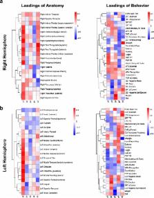
          Exercise was associated with increases in cortical thickness within the right pre and postcentral gyri. Other notable areas of increased thickness related to training were present in the left pre and postcentral gyri, left temporal pole, left superior temporal gyrus, and left parahippocampal gyrus. Further, we observed that compared to a separate cohort of healthy children, participants displayed multiple areas with a significantly thinner cortex prior to training and fewer differences following training, indicating amelioration of anatomical deficits. Partial least squares analysis (PLS) revealed specific patterns of relations between cortical thickness and various behavioral outcomes both after training and at baseline.

          Overall, our results indicate that exercise training in pediatric brain tumor patients treated with radiation has a beneficial impact on brain structure. We argue that exercise training should be incorporated into the development of neuro-rehabilitative treatments for long-term pediatric brain tumor survivors and other populations with acquired brain injury. ( ClinicalTrials.gov, NCT01944761)

          Highlights

          • Exercise training in pediatric brain tumor patients treated with radiation results in changes in brain structure

          • Exercise was associated with increased cortical thickness in several areas including motor and somatosensory cortex

          • Fewer differences between patients and healthy controls in cortical thickness were seen following exercise training

          • Specific patterns of relations between cortical thickness and behavior at a baseline and after exercise training were seen

          Related collections

          Most cited references63

          • Record: found
          • Abstract: found
          • Article: not found

          Partial Least Squares (PLS) methods for neuroimaging: a tutorial and review.

          Partial Least Squares (PLS) methods are particularly suited to the analysis of relationships between measures of brain activity and of behavior or experimental design. In neuroimaging, PLS refers to two related methods: (1) symmetric PLS or Partial Least Squares Correlation (PLSC), and (2) asymmetric PLS or Partial Least Squares Regression (PLSR). The most popular (by far) version of PLS for neuroimaging is PLSC. It exists in several varieties based on the type of data that are related to brain activity: behavior PLSC analyzes the relationship between brain activity and behavioral data, task PLSC analyzes how brain activity relates to pre-defined categories or experimental design, seed PLSC analyzes the pattern of connectivity between brain regions, and multi-block or multi-table PLSC integrates one or more of these varieties in a common analysis. PLSR, in contrast to PLSC, is a predictive technique which, typically, predicts behavior (or design) from brain activity. For both PLS methods, statistical inferences are implemented using cross-validation techniques to identify significant patterns of voxel activation. This paper presents both PLS methods and illustrates them with small numerical examples and typical applications in neuroimaging. Copyright © 2010 Elsevier Inc. All rights reserved.
            Bookmark
            • Record: found
            • Abstract: found
            • Article: not found

            Automatic 3D intersubject registration of MR volumetric data in standardized Talairach space.

            In both diagnostic and research applications, the interpretation of MR images of the human brain is facilitated when different data sets can be compared by visual inspection of equivalent anatomical planes. Quantitative analysis with predefined atlas templates often requires the initial alignment of atlas and image planes. Unfortunately, the axial planes acquired during separate scanning sessions are often different in their relative position and orientation, and these slices are not coplanar with those in the atlas. We have developed a completely automatic method to register a given volumetric data set with Talairach stereotaxic coordinate system. The registration method is based on multi-scale, three-dimensional (3D) cross-correlation with an average (n > 300) MR brain image volume aligned with the Talariach stereotaxic space. Once the data set is re-sampled by the transformation recovered by the algorithm, atlas slices can be directly superimposed on the corresponding slices of the re-sampled volume. the use of such a standardized space also allows the direct comparison, voxel to voxel, of two or more data sets brought into stereotaxic space. With use of a two-tailed Student t test for paired samples, there was no significant difference in the transformation parameters recovered by the automatic algorithm when compared with two manual landmark-based methods (p > 0.1 for all parameters except y-scale, where p > 0.05). Using root-mean-square difference between normalized voxel intensities as an unbiased measure of registration, we show that when estimated and averaged over 60 volumetric MR images in standard space, this measure was 30% lower for the automatic technique than the manual method, indicating better registrations. Likewise, the automatic method showed a 57% reduction in standard deviation, implying a more stable technique. The algorithm is able to recover the transformation even when data are missing from the top or bottom of the volume. We present a fully automatic registration method to map volumetric data into stereotaxic space that yields results comparable with those of manually based techniques. The method requires no manual identification of points or contours and therefore does not suffer the drawbacks involved in user intervention such as reproducibility and interobserver variability.
              Bookmark
              • Record: found
              • Abstract: found
              • Article: not found

              Sexual dimorphism of brain developmental trajectories during childhood and adolescence.

              Human total brain size is consistently reported to be approximately 8-10% larger in males, although consensus on regionally specific differences is weak. Here, in the largest longitudinal pediatric neuroimaging study reported to date (829 scans from 387 subjects, ages 3 to 27 years), we demonstrate the importance of examining size-by-age trajectories of brain development rather than group averages across broad age ranges when assessing sexual dimorphism. Using magnetic resonance imaging (MRI) we found robust male/female differences in the shapes of trajectories with total cerebral volume peaking at age 10.5 in females and 14.5 in males. White matter increases throughout this 24-year period with males having a steeper rate of increase during adolescence. Both cortical and subcortical gray matter trajectories follow an inverted U shaped path with peak sizes 1 to 2 years earlier in females. These sexually dimorphic trajectories confirm the importance of longitudinal data in studies of brain development and underline the need to consider sex matching in studies of brain development.
                Bookmark

                Author and article information

                Contributors
                Journal
                Neuroimage Clin
                Neuroimage Clin
                NeuroImage : Clinical
                Elsevier
                2213-1582
                05 March 2018
                2018
                05 March 2018
                : 18
                : 972-985
                Affiliations
                [a ]Neurosciences and Mental Health, Hospital for Sick Children, Toronto, Ontario, Canada
                [b ]Department of Pediatrics, McMaster University, Hamilton, Ontario, Canada
                [c ]Division of Hematology/Oncology, Hospital for Sick Children, Toronto, Ontario, Canada
                [d ]Department of Pediatrics, University of Toronto, Toronto, Ontario, Canada
                [e ]Diagnostic Imaging, Hospital for Sick Children, Toronto, Ontario, Canada
                [f ]Mouse Imaging Centre, Hospital for Sick Children, Toronto, Ontario, Canada
                [g ]Department of Medical Biophysics, University of Toronto, Toronto, Ontario, Canada
                [h ]Department of Psychology, University of Toronto, Toronto, Ontario, Canada
                Author notes
                [* ]Corresponding author at: Program in Neurosciences and Mental Health, The Hospital for Sick Children, 555 University Avenue, Toronto, Ontario M5G 1X8, Canada. donald.mabbott@ 123456sickkids.ca
                Article
                S2213-1582(18)30055-X
                10.1016/j.nicl.2018.02.021
                5987848
                29876282
                e702c934-f6bc-4611-9f3a-0889b408134d
                © 2018 The Authors

                This is an open access article under the CC BY-NC-ND license (http://creativecommons.org/licenses/by-nc-nd/4.0/).

                History
                : 11 August 2017
                : 23 December 2017
                : 21 February 2018
                Categories
                Regular Article

                brain recovery,cranial radiation,cortical thickness,exercise,neuroplasticity,pediatric brain tumor

                Comments

                Comment on this article