129
views
0
recommends
+1 Recommend
0 collections
    0
    shares
      • Record: found
      • Abstract: found
      • Article: found
      Is Open Access

      Porcine epidemic diarrhea virus (PEDV) co-infection induced chlamydial persistence/stress does not require viral replication

      research-article

      Read this article at

      Bookmark
          There is no author summary for this article yet. Authors can add summaries to their articles on ScienceOpen to make them more accessible to a non-specialist audience.

          Abstract

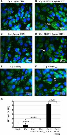
          Chlamydiae may exist at the site of infection in an alternative replicative form, called the aberrant body (AB). ABs are produced during a viable but non-infectious developmental state termed “persistence” or “chlamydial stress.” As persistent/stressed chlamydiae: (i) may contribute to chronic inflammation observed in diseases like trachoma; and (ii) are more resistant to current anti-chlamydial drugs of choice, it is critical to better understand this developmental stage. We previously demonstrated that porcine epidemic diarrhea virus (PEDV) co-infection induced Chlamydia pecorum persistence/stress in culture. One critical characteristic of persistence/stress is that the chlamydiae remain viable and can reenter the normal developmental cycle when the stressor is removed. Thus, we hypothesized that PEDV-induced persistence would be reversible if viral replication was inhibited. Therefore, we performed time course experiments in which Vero cells were C. pecorum/PEDV infected in the presence of cycloheximide (CHX), which inhibits viral but not chlamydial protein synthesis. CHX-exposure inhibited PEDV replication, but did not inhibit induction of C. pecorum persistence at 24 h post-PEDV infection, as indicated by AB formation and reduced production of infectious EBs. Interestingly, production of infectious EBs resumed when CHX-exposed, co-infected cells were incubated 48–72 h post-PEDV co-infection. These data demonstrate that PEDV co-infection-induced chlamydial persistence/stress is reversible and suggest that this induction (i) does not require viral replication in host cells; and (ii) does not require de novo host or viral protein synthesis. These data also suggest that viral binding and/or entry may be required for this effect. Because the PEDV host cell receptor (CD13 or aminopeptidase N) stimulates cellular signaling pathways in the absence of PEDV infection, we suspect that PEDV co-infection might alter CD13 function and induce the chlamydiae to enter the persistent state.

          Related collections

          Most cited references38

          • Record: found
          • Abstract: found
          • Article: not found

          Inactivation of the coronavirus that induces severe acute respiratory syndrome, SARS-CoV

          Severe acute respiratory syndrome (SARS) is a life-threatening disease caused by a novel coronavirus termed SARS-CoV. Due to the severity of this disease, the World Health Organization (WHO) recommends that manipulation of active viral cultures of SARS-CoV be performed in containment laboratories at biosafety level 3 (BSL3). The virus was inactivated by ultraviolet light (UV) at 254 nm, heat treatment of 65 °C or greater, alkaline (pH > 12) or acidic (pH < 3) conditions, formalin and glutaraldehyde treatments. We describe the kinetics of these efficient viral inactivation methods, which will allow research with SARS-CoV containing materials, that are rendered non-infectious, to be conducted at reduced safety levels.
            Bookmark
            • Record: found
            • Abstract: found
            • Article: not found

            Toll-like receptor and RIG-I-like receptor signaling.

            Toll-like receptors (TLRs) and RIG-I-like receptors (RLRs) constitute distinct families of pattern-recognition receptors that sense nucleic acids derived from viruses and trigger antiviral innate immune responses. TLR3, TLR7, and TLR9 are membrane proteins localized to the endosome that recognize viral double-stranded RNA, single-stranded RNA, and DNA, respectively, while RLRs, including RIG-I, Mda5, and LGP2, are cytoplasmic proteins that recognize viral RNA. Upon recognition of these nucleic acid species, TLRs and RLRs recruit specific intracellular adaptor proteins to initiate signaling pathways culminating in activation of NF-kappaB, MAP kinases, and IRFs that control the transcription of genes encoding type I interferon and other inflammatory cytokines, which are important for eliminating viruses. Here, we review recent insights into the signaling pathways initiated by TLR and RLR and their roles in innate and adaptive immune responses.
              Bookmark
              • Record: found
              • Abstract: found
              • Article: not found

              Multiple infections and the evolution of virulence.

              Infections that consist of multiple parasite strains or species are common in the wild and are a major public health concern. Theory suggests that these infections have a key influence on the evolution of infectious diseases and, more specifically, on virulence evolution. However, we still lack an overall vision of the empirical support for these predictions. We argue that within-host interactions between parasites largely determine how virulence evolves and that experimental data support model predictions. Then, we explore the main limitation of the experimental study of such 'mixed infections', which is that it draws conclusions on evolutionary outcomes from studies conducted at the individual level. We also discuss differences between coinfections caused by different strains of the same species or by different species. Overall, we argue that it is possible to make sense out of the complexity inherent to multiple infections and that experimental evolution settings may provide the best opportunity to further our understanding of virulence evolution. © 2013 Blackwell Publishing Ltd/CNRS.
                Bookmark

                Author and article information

                Journal
                Front Cell Infect Microbiol
                Front Cell Infect Microbiol
                Front. Cell. Infect. Microbiol.
                Frontiers in Cellular and Infection Microbiology
                Frontiers Media S.A.
                2235-2988
                09 January 2014
                13 March 2014
                2014
                : 4
                : 20
                Affiliations
                [1] 1Department of Biomedical Sciences, Quillen College of Medicine, East Tennessee State University Johnson City, TN, USA
                [2] 2Department of Pathobiology, Institute of Veterinary Pathology, University of Zurich Zurich, Switzerland
                Author notes

                Edited by: Jan Rupp, University of Lübeck, Germany

                Reviewed by: Thomas Rudel, University of Wuerzburg, Germany; Andreas Pospischil, University of Zurich, Switzerland

                *Correspondence: Nicole Borel, Vetsuisse Faculty, Institute of Veterinary Pathology, University of Zurich, Winterthurerstrasse 268, CH-8057 Zurich, Switzerland e-mail: n.borel@ 123456access.uzh.ch

                This article was submitted to the journal Frontiers in Cellular and Infection Microbiology.

                Article
                10.3389/fcimb.2014.00020
                3952398
                24660163
                a34653ab-7ce3-4c87-b4be-2706fd4b4704
                Copyright © 2014 Schoborg and Borel.

                This is an open-access article distributed under the terms of the Creative Commons Attribution License (CC BY). The use, distribution or reproduction in other forums is permitted, provided the original author(s) or licensor are credited and that the original publication in this journal is cited, in accordance with accepted academic practice. No use, distribution or reproduction is permitted which does not comply with these terms.

                History
                : 28 November 2013
                : 05 February 2014
                Page count
                Figures: 4, Tables: 0, Equations: 0, References: 48, Pages: 10, Words: 7659
                Categories
                Microbiology
                Original Research Article

                Infectious disease & Microbiology
                chlamydia pecorum,chlamydial persistence,chlamydial stress response,stressed chlamydiae,porcine epidemic diarrhea virus

                Comments

                Comment on this article