16
views
0
recommends
+1 Recommend
0 collections
    0
    shares
      • Record: found
      • Abstract: found
      • Article: found
      Is Open Access

      PEDV enters cells through clathrin-, caveolae-, and lipid raft-mediated endocytosis and traffics via the endo-/lysosome pathway

      research-article
      , , , ,
      Veterinary Research
      BioMed Central

      Read this article at

      Bookmark
          There is no author summary for this article yet. Authors can add summaries to their articles on ScienceOpen to make them more accessible to a non-specialist audience.

          Abstract

          With the emergence of highly pathogenic variant strains, porcine epidemic diarrhea virus (PEDV) has led to significant economic loss in the global swine industry. Many studies have described how coronaviruses enter cells, but information on PEDV invasion strategies remains insufficient. Given that the differences in gene sequences and pathogenicity between classical and mutant strains of PEDV may lead to diverse invasion mechanisms, this study focused on the cellular entry pathways and cellular transport of the PEDV GI and GII subtype strains in Vero cells and IPEC-J2 cells. We first characterized the kinetics of PEDV entry into cells and found that the highest invasion rate of PEDV was approximately 33% in the IPEC-J2 cells and approximately 100% in the Vero cells. To clarify the specific endocytic pathways, systematic research methods were used and showed that PEDV enters cells via the clathrin- and caveolae-mediated endocytosis pathways, in which dynamin II, clathrin heavy chain, Eps15, cholesterol, and caveolin-1 were indispensably involved. In addition, lipid raft extraction assay showed that PEDV can also enter cells through lipid raft-mediated endocytosis. To investigate the trafficking of internalized PEDV, we found that PEDV entry into cells relied on low pH and internalized virions reached lysosomes through the early endosome–late endosome–lysosome pathway. The results concretely revealed the entry mechanisms of PEDV and provided an insightful theoretical basis for the further understanding of PEDV pathogenesis and guidance for new targets of antiviral drugs.

          Related collections

          Most cited references53

          • Record: found
          • Abstract: found
          • Article: not found

          Porcine epidemic diarrhoea virus: a comprehensive review of molecular epidemiology, diagnosis, and vaccines

          The porcine epidemic diarrhoea virus (PEDV), a member of the Coronaviridae family, causes acute diarrhoea and dehydration in pigs. Although it was first identified in Europe, it has become increasingly problematic in many Asian countries, including Korea, China, Japan, the Philippines, and Thailand. The economic impacts of the PEDV are substantial, given that it results in significant morbidity and mortality in neonatal piglets and is associated with increased costs related to vaccination and disinfection. Recently, progress has been made in understanding the molecular epidemiology of PEDV, thereby leading to the development of new vaccines. In the current review, we first describe the molecular and genetic characteristics of the PEDV. Then we discuss its molecular epidemiology and diagnosis, what vaccines are available, and how PEDV can be treated.
            Bookmark
            • Record: found
            • Abstract: found
            • Article: found
            Is Open Access

            Outbreak of Porcine Epidemic Diarrhea in Suckling Piglets, China

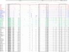
            To the Editor: Beginning in October 2010, porcine epidemic diarrhea (PED), caused by a coronaviral infection affecting pigs, emerged in China in an outbreak characterized by high mortality rates among suckling piglets. The outbreak overwhelmed >10 provinces in southern China, and >1,000,000 piglets died. This outbreak was distinguished by ≈100% illness among piglets after birth (predominantly within 7 days and sometimes within only a few hours) and death rates of 80%–100% (Technical Appendix Table 1). Few sows or boars showed any clinical signs during the outbreak, which is not consistent with a recent report from Thailand ( 1 ). In that outbreak during late 2007, pigs of all ages were affected, exhibiting different degrees of diarrhea and no appetite. We characterized the genetic variation of the PED virus (PEDV) that caused a large-scale outbreak in China during 2010–2011 and compared it with viruses in other outbreaks. We also report a possible novel transmission pathway for PEDV. A total of 177 samples (intestine, stool, and maternal milk) were collected from pigs from different farms who had diarrhea; 100% of farms had >1 porcine sample positive for PEDV. A total of 125/177 porcine samples were confirmed as positive for PEDV by reverse transcription PCR using primers as described ( 2 ). PEDV was detected in 105 (82.0%) of 128 fecal samples and 20 (40.8%) of 49 sow milk samples. Piglets infected with PEDV showed mild hemorrhage, undigested curdled milk in the stomach, and thin-walled intestines with severe mucosal atrophy and foamy fluid (data not shown). The spike (S) gene of the family Coronaviridae has a high degree of variation and can induce neutralizing antibody ( 3 ). Reverse transcription PCR products of the 651-bp partial S gene of PEDV and the deduced amino acid sequences were aligned by using ClustalW (www.genome.jp/tools/clustalw), and a neighbor-joining tree with 1,000 bootstraps was constructed. Sequences of the S genes from this outbreak were 99.1%–100.0% homologous and had 88.7%–98.9% nt identity with all reference strains (Technical Appendix Table 2), 98.5%–98.9% with Thailand strains, and 94.5%–95.1% with vaccine strain CV777. The partial S gene deduced amino acid sequences were compared and also showed a high degree of homology (98.0%–100.0%); they had 85.3%–98.7% identity with all reference strains listed in Technical Appendix Table 2, 98.0%–98.7% with Thailand strains, and 93.3%–94.7% with vaccine strain CV777 (data not shown). Phylogenetic analysis indicated that the PEDV in the China outbreak was different from foreign and other domestic strains on the basis of the reported partial S gene sequences. All new strains were clustered in the same branch, close to the cluster of Thailand strains, and far from the cluster of vaccine strain CV777 (Figure). Figure Phylogenetic tree constructed by using the neighbor-joining method based on the 9 porcine epidemic diarrhea virus (PEDV) sequences identified in a study of porcine epidemic diarrhea in China. Partially amplified spike genes of the PEDV isolates plus 18 PEDV sequences downloaded from GenBank were compared. Sequences included in each cluster are listed in Technical Appendix Table 3. Strains from Thailand and China and the CV777 vaccine strain are indicated. Scale bar indicates nucleotide substitutions per site. In the China outbreak, PEDV caused severe diarrheal disease in piglets; heavy economic losses in many provinces resulted, despite use of commercial vaccines (inactivated transmissible gastroenteritis [TGEV H] and porcine epidemic diarrhea [CV777]). To determine why the vaccines showed poor efficacy, we investigated evolution of the virus. Comparison of amino acid sequences from isolates from the outbreak and from the CV777 vaccine strain showed 9 amino acid mutations of fragments containing major hydrophilic regions: 16 (L→H), 18 (S→G), 22 (V→I), 44 (T→S), 89 (G→S), 100 (A→E), 107 (L→F), 130 (I→V) and 160 (I→F) (Technical Appendix Figure, panel A). Three of these 9 mutations were at positions 16, 18, and 22 in the isolates from China; they influenced the hydrophobicity of the S protein as compared with that for CV777 (Technical Appendix Figure, panel B). Phylogenic analysis showed that strain CV777 did not cluster with current common strains and showed considerable genetic distance from them. Isolates in the outbreak in China had only a minor nucleotide sequence variation from the Thailand isolates, indicating that the virus has a high genetic relatedness to the Southeast Asia strain. However, previous studies showed that isolates from Europe, South Korea, and China were serologically identical to the prototype CV777 strain ( 1 , 4 ). To our knowledge, fecal–oral transmission is probably the main or only route of PEDV transmission ( 5 – 7 ). In our study, if a fecal sample from a sick piglet was found to be positive for PEDV, we also collected and studied milk from its mother. These results showed that PEDV was present in sow milk (Technical Appendix Table 3), but the detection rate was lower for these samples (40.8%) than for the fecal samples (82.0%). On the basis of these results, we hypothesize that sow milk could represent a possible (and potentially major) route for the vertical transmission of PEDV from sow to suckling piglet. This hypothesis could be indirectly verified by our field observation that piglet death rates decreased as a result of fostering (data not shown). Our findings show that PEDV was identified not only in fecal samples from sick piglets, as expected, but also in the milk of the sow, which suggests vertical transmission of the virus. Supplementary Material Technical Appendix Current farms status in this study, China.
              Bookmark
              • Record: found
              • Abstract: found
              • Article: found
              Is Open Access

              Porcine epidemic diarrhea virus: An emerging and re-emerging epizootic swine virus

              The enteric disease of swine recognized in the early 1970s in Europe was initially described as “epidemic viral diarrhea” and is now termed “porcine epidemic diarrhea (PED)”. The coronavirus referred to as PED virus (PEDV) was determined to be the etiologic agent of this disease in the late 1970s. Since then the disease has been reported in Europe and Asia, but the most severe outbreaks have occurred predominantly in Asian swine-producing countries. Most recently, PED first emerged in early 2013 in the United States that caused high morbidity and mortality associated with PED, remarkably affecting US pig production, and spread further to Canada and Mexico. Soon thereafter, large-scale PED epidemics recurred through the pork industry in South Korea, Japan, and Taiwan. These recent outbreaks and global re-emergence of PED require urgent attention and deeper understanding of PEDV biology and pathogenic mechanisms. This paper highlights the current knowledge of molecular epidemiology, diagnosis, and pathogenesis of PEDV, as well as prevention and control measures against PEDV infection. More information about the virus and the disease is still necessary for the development of effective vaccines and control strategies. It is hoped that this review will stimulate further basic and applied studies and encourage collaboration among producers, researchers, and swine veterinarians to provide answers that improve our understanding of PEDV and PED in an effort to eliminate this economically significant viral disease, which emerged or re-emerged worldwide.
                Bookmark

                Author and article information

                Contributors
                weixiaona_na@126.com
                1123856877@qq.com
                1942904309@qq.com
                xuecy@mail.sysu.edu.cn
                caoych@mail.sysu.edu.cn
                Journal
                Vet Res
                Vet. Res
                Veterinary Research
                BioMed Central (London )
                0928-4249
                1297-9716
                10 February 2020
                10 February 2020
                2020
                : 51
                : 10
                Affiliations
                GRID grid.12981.33, ISNI 0000 0001 2360 039X, State Key Laboratory of Biocontrol, School of Life Sciences, , Sun Yat-sen University, ; Guangzhou, 510006 People’s Republic of China
                Author information
                http://orcid.org/0000-0002-0311-5474
                Article
                739
                10.1186/s13567-020-0739-7
                7011528
                32041637
                d4b6d41b-f540-4dfb-8baa-2b2ac167e041
                © The Author(s) 2020

                Open AccessThis article is licensed under a Creative Commons Attribution 4.0 International License, which permits use, sharing, adaptation, distribution and reproduction in any medium or format, as long as you give appropriate credit to the original author(s) and the source, provide a link to the Creative Commons licence, and indicate if changes were made. The images or other third party material in this article are included in the article's Creative Commons licence, unless indicated otherwise in a credit line to the material. If material is not included in the article's Creative Commons licence and your intended use is not permitted by statutory regulation or exceeds the permitted use, you will need to obtain permission directly from the copyright holder. To view a copy of this licence, visit http://creativecommons.org/licenses/by/4.0/. The Creative Commons Public Domain Dedication waiver ( http://creativecommons.org/publicdomain/zero/1.0/) applies to the data made available in this article, unless otherwise stated in a credit line to the data.

                History
                : 5 October 2019
                : 7 January 2020
                Funding
                Funded by: National Key Research and Development Program of China
                Award ID: 2016YFD0500101
                Award Recipient :
                Categories
                Research Article
                Custom metadata
                © L'Institut National de Recherche en Agriculture, Alimentation et Environnement (INRAE) 2020

                Veterinary medicine
                Veterinary medicine

                Comments

                Comment on this article