35
views
0
recommends
+1 Recommend
0 collections
    0
    shares
      • Record: found
      • Abstract: found
      • Article: found
      Is Open Access

      Withaferin A Induces Proteasome Inhibition, Endoplasmic Reticulum Stress, the Heat Shock Response and Acquisition of Thermotolerance

      research-article
      , , *
      PLoS ONE
      Public Library of Science

      Read this article at

      Bookmark
          There is no author summary for this article yet. Authors can add summaries to their articles on ScienceOpen to make them more accessible to a non-specialist audience.

          Abstract

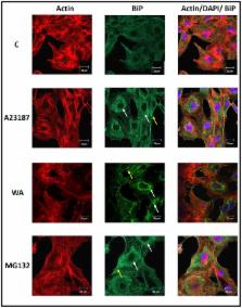
          In the present study, withaferin A (WA), a steroidal lactone with anti-inflammatory and anti-tumor properties, inhibited proteasome activity and induced endoplasmic reticulum (ER) and cytoplasmic HSP accumulation in Xenopus laevis A6 kidney epithelial cells. Proteasomal inhibition by WA was indicated by an accumulation of ubiquitinated protein and a decrease in chymotrypsin-like activity. Additionally, immunoblot analysis revealed that treatment of cells with WA induced the accumulation of HSPs including ER chaperones, BiP and GRP94, as well as cytoplasmic/nuclear HSPs, HSP70 and HSP30. Furthermore, WA-induced an increase in the relative levels of the protein kinase, Akt, while the levels of actin were unchanged compared to control. Northern blot experiments determined that WA induced an accumulation in bip, hsp70 and hsp30 mRNA but not eIF-1α mRNA. Interestingly, WA acted synergistically with mild heat shock to enhance HSP70 and HSP30 accumulation to a greater extent than the sum of both stressors individually. This latter phenomenon was not observed with BiP or GRP94. Immunocytochemical analysis indicated that WA-induced BiP accumulation occurred mainly in the perinuclear region in a punctate pattern, while HSP30 accumulation occurred primarily in a granular pattern in the cytoplasm with some staining in the nucleus. Prolonged exposure to WA resulted in disorganization of the F-actin cytoskeleton as well as the production of relatively large HSP30 staining structures that co-localized with F-actin. Finally, prior exposure of cells to WA treatment, which induced the accumulation of HSPs conferred a state of thermal protection since it protected the F-actin cytoskeleton against a subsequent cytotoxic thermal challenge.

          Related collections

          Most cited references62

          • Record: found
          • Abstract: found
          • Article: not found

          Aggresomes: A Cellular Response to Misfolded Proteins

          Intracellular deposition of misfolded protein aggregates into ubiquitin-rich cytoplasmic inclusions is linked to the pathogenesis of many diseases. Why these aggregates form despite the existence of cellular machinery to recognize and degrade misfolded protein and how they are delivered to cytoplasmic inclusions are not known. We have investigated the intracellular fate of cystic fibrosis transmembrane conductance regulator (CFTR), an inefficiently folded integral membrane protein which is degraded by the cytoplasmic ubiquitin-proteasome pathway. Overexpression or inhibition of proteasome activity in transfected human embryonic kidney or Chinese hamster ovary cells led to the accumulation of stable, high molecular weight, detergent-insoluble, multiubiquitinated forms of CFTR. Using immunofluorescence and transmission electron microscopy with immunogold labeling, we demonstrate that undegraded CFTR molecules accumulate at a distinct pericentriolar structure which we have termed the aggresome. Aggresome formation is accompanied by redistribution of the intermediate filament protein vimentin to form a cage surrounding a pericentriolar core of aggregated, ubiquitinated protein. Disruption of microtubules blocks the formation of aggresomes. Similarly, inhibition of proteasome function also prevented the degradation of unassembled presenilin-1 molecules leading to their aggregation and deposition in aggresomes. These data lead us to propose that aggresome formation is a general response of cells which occurs when the capacity of the proteasome is exceeded by the production of aggregation-prone misfolded proteins.
            Bookmark
            • Record: found
            • Abstract: found
            • Article: not found

            Endoplasmic reticulum stress in liver disease.

            The unfolded protein response (UPR) is activated upon the accumulation of misfolded proteins in the endoplasmic reticulum (ER) that are sensed by the binding immunoglobulin protein (BiP)/glucose-regulated protein 78 (GRP78). The accumulation of unfolded proteins sequesters BiP so it dissociates from three ER-transmembrane transducers leading to their activation. These transducers are inositol requiring (IRE) 1α, PKR-like ER kinase (PERK), and activating transcription factor (ATF) 6α. PERK phosphorylates eukaryotic initiation factor 2 alpha (eIF2α) resulting in global mRNA translation attenuation, and concurrently selectively increases the translation of several mRNAs, including the transcription factor ATF4, and its downstream target CHOP. IRE1α has kinase and endoribonuclease (RNase) activities. IRE1α autophosphorylation activates the RNase activity to splice XBP1 mRNA, to produce the active transcription factor sXBP1. IRE1α activation also recruits and activates the stress kinase JNK. ATF6α transits to the Golgi compartment where it is cleaved by intramembrane proteolysis to generate a soluble active transcription factor. These UPR pathways act in concert to increase ER content, expand the ER protein folding capacity, degrade misfolded proteins, and reduce the load of new proteins entering the ER. All of these are geared toward adaptation to resolve the protein folding defect. Faced with persistent ER stress, adaptation starts to fail and apoptosis occurs, possibly mediated through calcium perturbations, reactive oxygen species, and the proapoptotic transcription factor CHOP. The UPR is activated in several liver diseases; including obesity associated fatty liver disease, viral hepatitis, and alcohol-induced liver injury, all of which are associated with steatosis, raising the possibility that ER stress-dependent alteration in lipid homeostasis is the mechanism that underlies the steatosis. Hepatocyte apoptosis is a pathogenic event in several liver diseases, and may be linked to unresolved ER stress. If this is true, restoration of ER homeostasis prior to ER stress-induced cell death may provide a therapeutic rationale in these diseases. Herein we discuss each branch of the UPR and how they may impact hepatocyte function in different pathologic states. Copyright © 2010 European Association for the Study of the Liver. Published by Elsevier B.V. All rights reserved.
              Bookmark
              • Record: found
              • Abstract: found
              • Article: not found

              Proteotoxic stress and inducible chaperone networks in neurodegenerative disease and aging.

              The long-term health of the cell is inextricably linked to protein quality control. Under optimal conditions this is accomplished by protein homeostasis, a highly complex network of molecular interactions that balances protein biosynthesis, folding, translocation, assembly/disassembly, and clearance. This review will examine the consequences of an imbalance in homeostasis on the flux of misfolded proteins that, if unattended, can result in severe molecular damage to the cell. Adaptation and survival requires the ability to sense damaged proteins and to coordinate the activities of protective stress response pathways and chaperone networks. Yet, despite the abundance and apparent capacity of chaperones and other components of homeostasis to restore folding equilibrium, the cell appears poorly adapted for chronic proteotoxic stress when conformationally challenged aggregation-prone proteins are expressed in cancer, metabolic disease, and neurodegenerative disease. The decline in biosynthetic and repair activities that compromises the integrity of the proteome is influenced strongly by genes that control aging, thus linking stress and protein homeostasis with the health and life span of the organism.
                Bookmark

                Author and article information

                Contributors
                Role: Editor
                Journal
                PLoS One
                PLoS ONE
                plos
                plosone
                PLoS ONE
                Public Library of Science (San Francisco, USA )
                1932-6203
                2012
                30 November 2012
                : 7
                : 11
                : e50547
                Affiliations
                [1]Department of Biology, University of Waterloo, Waterloo, Ontario, Canada
                Virginia Commonwealth University, United States of America
                Author notes

                Competing Interests: The authors have declared that no competing interests exist.

                Conceived and designed the experiments: JJH SK AWR. Performed the experiments: SK AWR. Analyzed the data: JJH SK AWR. Contributed reagents/materials/analysis tools: JJH. Wrote the paper: JJH SK AWR.

                Article
                PONE-D-12-18902
                10.1371/journal.pone.0050547
                3511540
                23226310
                031b95ae-7b81-43df-bf41-02bc245a13db
                Copyright @ 2012

                This is an open-access article distributed under the terms of the Creative Commons Attribution License, which permits unrestricted use, distribution, and reproduction in any medium, provided the original author and source are credited.

                History
                : 29 June 2012
                : 25 October 2012
                Page count
                Pages: 14
                Funding
                This research was supported by Natural Science and Engineering Research Council (NSERC) grants to J.J.H. who is also the recipient of a Canada Research Chair in Stress Protein Gene Research. S.K. is the recipient of an NSERC graduate scholarship. The funders had no role in study design, data collection and analysis, decision to publish, or preparation of the manuscript.
                Categories
                Research Article
                Biology
                Anatomy and Physiology
                Cell Physiology
                Biochemistry
                Proteins
                Chaperone Proteins
                Cytoskeletal Proteins
                Genetics
                Molecular Genetics
                Gene Regulation
                Gene Expression
                Model Organisms
                Animal Models
                Xenopus Laevis
                Molecular Cell Biology
                Cellular Stress Responses
                Medicine
                Drugs and Devices
                Clinical Pharmacology

                Uncategorized
                Uncategorized

                Comments

                Comment on this article