14
views
0
recommends
+1 Recommend
0 collections
    0
    shares
      • Record: found
      • Abstract: found
      • Article: found
      Is Open Access

      Comparison of Clinical and Biomechanical Outcomes of Small Incision Lenticule Extraction With 120- and 140-µm Cap Thickness

      research-article

      Read this article at

      Bookmark
          There is no author summary for this article yet. Authors can add summaries to their articles on ScienceOpen to make them more accessible to a non-specialist audience.

          Abstract

          Purpose

          The purpose of this study was to compare the clinical outcomes and corneal biomechanical changes between 120-µm and 140-µm cap thickness after small incision lenticule extraction (SMILE).

          Methods

          This prospective study included 150 eyes (150 patients: 91 eyes in the 120-µm group, and 59 eyes in the 140-µm group) who underwent SMILE. Enhanced correction nomograms were applied for patients according to cap thickness. Clinical outcomes, including visual acuity, refraction, and corneal wavefront aberrations, were compared between the two groups. Corneal biomechanics were evaluated using the Corvis ST (Oculus, Wetzlar, Germany).

          Results

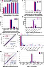
          The mean uncorrected-distance visual acuity, safety and efficacy indices, and refractive predictability were comparable in the 120-µm and 140-µm groups after SMILE. The postoperative total corneal root mean square higher-order aberrations (HOAs) and spherical aberrations was 0.48 ± 0.31 and 0.26 ± 0.10 in the 120-µm group, and 0.53 ± 0.16 and 0.34 ± 0.13 in the 140-µm group, which showed significant differences between the two groups ( P = 0.027, and <0.001, respectively). Although corneal stiffness decreased after SMILE in both groups, the changes in the deformation amplitude ratio were significantly higher in the 140-µm group than in the 120-µm group ( P = 0.022).

          Conclusions

          SMILE with 120-µm and 140-µm cap thickness provided excellent predictable outcomes according to our enhanced correction nomogram. The amount of tissue removal required to achieve the same amount of refractive correction was greater in the thicker cap group. The induction of corneal HOAs and weakening of corneal biomechanics were less pronounced in the thin-cap group, which may be associated with the thinner cap, lesser lenticule thickness, or thicker residual stromal bed.

          Translational Relevance

          Although SMILE with different cap thickness was effective, thicker lenticule thickness in the thick-cap group may be associated with induction of HOAs, and corneal stiffness changes.

          Related collections

          Most cited references41

          • Record: found
          • Abstract: found
          • Article: not found

          Progress in corneal wound healing.

          Corneal wound healing is a complex process involving cell death, migration, proliferation, differentiation, and extracellular matrix remodeling. Many similarities are observed in the healing processes of corneal epithelial, stromal and endothelial cells, as well as cell-specific differences. Corneal epithelial healing largely depends on limbal stem cells and remodeling of the basement membrane. During stromal healing, keratocytes get transformed to motile and contractile myofibroblasts largely due to activation of transforming growth factor-β (TGF-β) system. Endothelial cells heal mostly by migration and spreading, with cell proliferation playing a secondary role. In the last decade, many aspects of wound healing process in different parts of the cornea have been elucidated, and some new therapeutic approaches have emerged. The concept of limbal stem cells received rigorous experimental corroboration, with new markers uncovered and new treatment options including gene and microRNA therapy tested in experimental systems. Transplantation of limbal stem cell-enriched cultures for efficient re-epithelialization in stem cell deficiency and corneal injuries has become reality in clinical setting. Mediators and course of events during stromal healing have been detailed, and new treatment regimens including gene (decorin) and stem cell therapy for excessive healing have been designed. This is a very important advance given the popularity of various refractive surgeries entailing stromal wound healing. Successful surgical ways of replacing the diseased endothelium have been clinically tested, and new approaches to accelerate endothelial healing and suppress endothelial-mesenchymal transformation have been proposed including Rho kinase (ROCK) inhibitor eye drops and gene therapy to activate TGF-β inhibitor SMAD7. Promising new technologies with potential for corneal wound healing manipulation including microRNA, induced pluripotent stem cells to generate corneal epithelium, and nanocarriers for corneal drug delivery are discussed. Attention is also paid to problems in wound healing understanding and treatment, such as lack of specific epithelial stem cell markers, reliable identification of stem cells, efficient prevention of haze and stromal scar formation, lack of data on wound regulating microRNAs in keratocytes and endothelial cells, as well as virtual lack of targeted systems for drug and gene delivery to select corneal cells.
            Bookmark
            • Record: found
            • Abstract: found
            • Article: not found

            Results of small incision lenticule extraction: All-in-one femtosecond laser refractive surgery.

            To report the clinical results of small incision lenticule extraction to correct refractive errors using a femtosecond laser to refine the femtosecond lenticule extraction technique. Private laser center, Vadodara, India. Prospective clinical study. The VisuMax femtosecond laser system was used to perform small incision lenticule extraction to treat refractive errors. The laser was used to cut a refractive lenticule intrastromally to correct myopia and myopic astigmatism. The lenticule was then extracted from the stroma through a 3.0 to 5.0 mm incision. Outcome measures were corrected distance visual acuity (CDVA), uncorrected distance visual acuity (UDVA), and manifest refraction during 6 months of follow-up. Corneal topography and ocular wavefront aberrations were also measured. The study enrolled 51 eyes of 41 patients. The mean spherical equivalent was -4.87 diopters (D) ± 2.16 (SD) preoperatively and +0.03 ± 0.30 D 6 months postoperatively. Refractive stability was achieved within 1 month (P<.01). Six months after surgery, 79% of all full-correction cases had a UDVA of 20/25 or better. The 6-month postoperative CDVA was the same as or better than the preoperative CDVA in 95% of eyes. Two eyes lost 1 line of CDVA. All-in-one femtosecond refractive correction using a small incision technique was safe, predictable, and effective in treating myopia and myopic astigmatism. No author has a financial or proprietary interest in any material or method mentioned. Additional disclosure is found in the footnotes. Copyright © 2011 ASCRS and ESCRS. Published by Elsevier Inc. All rights reserved.
              Bookmark
              • Record: found
              • Abstract: found
              • Article: not found

              Risk factors and prognosis for corneal ectasia after LASIK.

              To review cases of corneal ectasia after laser in situ keratomileusis (LASIK), identify preoperative risk factors, and evaluate methods and success rates of visual rehabilitation for these cases. Retrospective nonrandomized comparative trial. Ten eyes from seven patients identified as developing corneal ectasia after LASIK, 33 previously reported ectasia cases, and two control groups with uneventful LASIK and normal postoperative courses: 100 consecutive cases (first control group), and 100 consecutive cases with high myopia (> 8 diopters [D]) preoperatively (second control group). Retrospective review of preoperative and postoperative data for each case compared with that of previously reported cases and cases with uneventful postoperative courses. Preoperative refraction, topographic features, residual stromal bed thickness (RSB), time to the development of ectasia, number of enhancements, final best-corrected visual acuity (BCVA), and method of final correction. Length of follow-up averaged 23.4 months (range, 6-48 months) after LASIK. Mean time to the development of ectasia averaged 16.3 months (range, 1-45 months). Preoperative refraction averaged -8.69 D compared with -5.37 D for the first control group (P = 0.005). Preoperatively, 88% of ectasia cases met criteria for forme fruste keratoconus, compared with 2% of the first control group (P < 0.0000001) and 4% of the second control group (P = 0.0000001). Seven eyes (70%) had RSB <250 microm, as did 16% of eyes in the first control group and 46% of the second control group. The mean RSB for ectasia cases (222.8 microm) was significantly less than that for the first control group (293.6 micro m, P = 0.0004) and the second control group (256.5 microm; P = 0.04). Seven eyes (70%) had enhancements. Only 10% of eyes lost more than one line of BCVA, and all patients eventually achieved corrected vision of 20/30 or better. One case required penetrating keratoplasty (10%), while all others required rigid gas-permeable contact lenses for correction. Significant risk factors for the development of ectasia after LASIK include high myopia, forme fruste keratoconus, and low RSB. All patients had at least one risk factor other than high myopia, and significant differences remained even when controlling for myopia. Multiple enhancements were common among affected cases, but their causative role remains unknown. We did not identify any patients who developed ectasia without recognizable preoperative risk factors.
                Bookmark

                Author and article information

                Journal
                Transl Vis Sci Technol
                Transl Vis Sci Technol
                tvst
                TVST
                Translational Vision Science & Technology
                The Association for Research in Vision and Ophthalmology
                2164-2591
                14 July 2021
                July 2021
                : 10
                : 8
                : 15
                Affiliations
                [1 ]The Institute of Vision Research, Department of Ophthalmology, Yonsei University College of Medicine, Seoul, Republic of Korea
                [2 ]Corneal Dystrophy Research Institute, Department of Ophthalmology, Yonsei University College of Medicine, Seoul, Republic of Korea
                [3 ]Eyereum Eye Clinic, Seoul, Republic of Korea
                [4 ]Department of Ophthalmology & Visual Sciences and Department of Biomedical Engineering, The Ohio State University, Columbus, OH, USA
                [5 ]Department of Ophthalmology, University of Ulsan College of Medicine, Seoul, Republic of Korea
                [6 ]Saevit Eye Hospital, Goyang-Si, Gyeonggi-Do, Republic of Korea
                Author notes
                [* ] Correspondence: Tae-im Kim, Department of Ophthalmology, Yonsei University College of Medicine, 50-1 Yonsei-ro, Seodaemungu, Seoul 03722, Republic of Korea. e-mail: tikim@ 123456yuhs.ac
                Article
                TVST-21-3322
                10.1167/tvst.10.8.15
                8288062
                34259803
                f1728acf-28d1-4fe3-b233-944ab1f3ee9b
                Copyright 2021 The Authors

                This work is licensed under a Creative Commons Attribution-NonCommercial-NoDerivatives 4.0 International License.

                History
                : 01 June 2021
                : 01 January 2021
                Page count
                Pages: 12
                Categories
                Article
                Article

                corneal biomechanics,smile,cap thickness,visual outcome
                corneal biomechanics, smile, cap thickness, visual outcome

                Comments

                Comment on this article