42
views
0
recommends
+1 Recommend
0 collections
    4
    shares
      • Record: found
      • Abstract: found
      • Article: not found

      Connexin 43 connexon to gap junction transition is regulated by zonula occludens-1

      research-article

      Read this article at

      ScienceOpenPublisherPMC
      Bookmark
          There is no author summary for this article yet. Authors can add summaries to their articles on ScienceOpen to make them more accessible to a non-specialist audience.

          Abstract

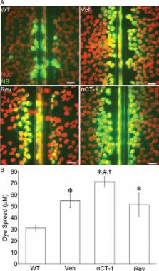
          Cx43 gap junctions (GJs) are integral to the function of the mammalian heart. It is shown that ZO-1 dynamically regulates the transition between Cx43 connexons and GJ intercellular channels, determining the balance of connexon-mediated cell permeability to GJ communication. Importantly, a novel domain proximal to GJs is identified—the perinexus.

          Abstract

          Connexin 43 (Cx43) is a gap junction (GJ) protein widely expressed in mammalian tissues that mediates cell-to-cell coupling. Intercellular channels comprising GJ aggregates form from docking of paired connexons, with one each contributed by apposing cells. Zonula occludens-1 (ZO-1) binds the carboxy terminus of Cx43, and we have previously shown that inhibition of the Cx43/ZO-1 interaction increases GJ size by 48 h. Here we demonstrated that increases in GJ aggregation occur within 2 h (∼Cx43 half-life) following disruption of Cx43/ZO-1. Immunoprecipitation and Duolink protein–protein interaction assays indicated that inhibition targets ZO-1 binding with Cx43 in GJs as well as connexons in an adjacent domain that we term the “perinexus.” Consistent with GJ size increases being matched by decreases in connexons, inhibition of Cx43/ZO-1 reduced the extent of perinexal interaction, increased the proportion of connexons docked in GJs relative to undocked connexons in the plasma membrane, and increased GJ intercellular communication while concomitantly decreasing hemichannel-mediated membrane permeance in contacting, but not noncontacting, cells. ZO-1 small interfering RNA and overexpression experiments verified that loss and gain of ZO-1 function govern the transition of connexons into GJs. It is concluded that ZO-1 regulates the rate of undocked connexon aggregation into GJs, enabling dynamic partitioning of Cx43 channel function between junctional and proximal nonjunctional domains of plasma membrane.

          Related collections

          Most cited references61

          • Record: found
          • Abstract: found
          • Article: found
          Is Open Access

          Remodelling of gap junctions and connexin expression in diseased myocardium

          Gap junctions form the cell-to-cell pathways for propagation of the precisely orchestrated patterns of current flow that govern the regular rhythm of the healthy heart. As in most tissues and organs, multiple connexin types are expressed in the heart: connexin43 (Cx43), Cx40 and Cx45 are found in distinctive combinations and relative quantities in different, functionally-specialized subsets of cardiac myocyte. Mutations in genes that encode connexins have only rarely been identified as being a cause of human cardiac disease, but remodelling of connexin expression and gap junction organization are well documented in acquired adult heart disease, notably ischaemic heart disease and heart failure. Remodelling may take the form of alterations in (i) the distribution of gap junctions and (ii) the amount and type of connexins expressed. Heterogeneous reduction in Cx43 expression and disordering in gap junction distribution feature in human ventricular disease and correlate with electrophysiologically identified arrhythmic changes and contractile dysfunction in animal models. Disease-related alterations in Cx45 and Cx40 expression have also been reported, and some of the functional implications of these are beginning to emerge. Apart from ventricular disease, various features of gap junction organization and connexin expression have been implicated in the initiation and persistence of the most common form of atrial arrhythmia, atrial fibrillation, though the disparate findings in this area remain to be clarified. Other major tasks ahead focus on the Purkinje/working ventricular myocyte interface and its role in normal and abnormal impulse propagation, connexin-interacting proteins and their regulatory functions, and on defining the precise functional properties conferred by the distinctive connexin co-expression patterns of different myocyte types in health and disease.
            Bookmark
            • Record: found
            • Abstract: found
            • Article: not found

            The interaction between Stargazin and PSD-95 regulates AMPA receptor surface trafficking.

            Accumulation of AMPA receptors at synapses is a fundamental feature of glutamatergic synaptic transmission. Stargazin, a member of the TARP family, is an AMPAR auxiliary subunit allowing interaction of the receptor with scaffold proteins of the postsynaptic density, such as PSD-95. How PSD-95 and Stargazin regulate AMPAR number in synaptic membranes remains elusive. We show, using single quantum dot and FRAP imaging in live hippocampal neurons, that exchange of AMPAR by lateral diffusion between extrasynaptic and synaptic sites mostly depends on the interaction of Stargazin with PSD-95 and not upon the GluR2 AMPAR subunit C terminus. Disruption of interactions between Stargazin and PSD-95 strongly increases AMPAR surface diffusion, preventing AMPAR accumulation at postsynaptic sites. Furthermore, AMPARs and Stargazin diffuse as complexes in and out synapses. These results propose a model in which the Stargazin-PSD-95 interaction plays a key role to trap and transiently stabilize diffusing AMPARs in the postsynaptic density.
              Bookmark
              • Record: found
              • Abstract: found
              • Article: not found

              Gating and regulation of connexin 43 (Cx43) hemichannels.

              Connexin 43 (Cx43) nonjunctional or "unapposed" hemichannels can open under physiological or pathological conditions. We characterize hemichannels comprised of Cx43 or Cx43-EGFP (Cx43 with enhanced GFP fused to the C terminus) expressed in HeLa cells. Channel opening was induced at potentials greater than +60 mV. Open probability appeared to be very low. No comparable opening was detected in the parental, nontransfected HeLa cells. Conductance of fully open single hemichannels was approximately 220 pS, which is approximately double that of Cx43 cell-cell channels. Cx43 hemichannels exhibited two types of gating: fast transitions ( 5 ms) between either open state and the fully closed state. Cx43-EGFP hemichannels exhibited only slow transitions (>5 ms) between closed and fully open states. These properties resemble those of the corresponding Cx43 and Cx43-EGFP cell-cell channels. Cx43 with EGFP on the N terminus (EGFP-Cx43) inserted into the surface and formed plaques but did not form hemichannels or cell-cell channels. Hemichannel blockers, 18beta-glycyrrhetinic acid or La3+, blocked depolarization-induced currents. Uptake of ethidium bromide (i) was faster in Cx43 and Cx43-EGFP than parental and EGFP-Cx43 cells, (ii) was directly correlated with Cx43-EGFP expression, (iii) was reduced by hemichannel blockers, and (iv) occurred at the same low rate in EGFP-Cx43 and parental cells. Although hemichannel opening was not detected electrophysiologically at the resting potential, infrequent or brief opening could account for ethidium bromide uptake. Opening of Cx43 hemichannels may mediate normal signaling or be deleterious.
                Bookmark

                Author and article information

                Contributors
                Role: Monitoring Editor
                Journal
                Mol Biol Cell
                molbiolcell
                mbc
                Mol. Bio. Cell
                Molecular Biology of the Cell
                The American Society for Cell Biology
                1059-1524
                1939-4586
                01 May 2011
                : 22
                : 9
                : 1516-1528
                Affiliations
                [1]Department of Regenerative Medicine and Cell Biology, Medical University of South Carolina, Charleston, SC 29425
                Emory University
                Author notes
                Address correspondence to: Robert G. Gourdie ( gourdier@ 123456musc.edu ).
                Article
                E10-06-0548
                10.1091/mbc.E10-06-0548
                3084674
                21411628
                edc48c39-54d4-4377-9f31-166e8ffc9ba7
                © 2011 Rhett et al. This article is distributed by The American Society for Cell Biology under license from the author(s). Two months after publication it is available to the public under an Attribution–Noncommercial–Share Alike 3.0 Unported Creative Commons License ( http://creativecommons.org/licenses/by-nc-sa/3.0).

                “ASCB®,“ “The American Society for Cell Biology®,” and “Molecular Biology of the Cell®” are registered trademarks of The American Society of Cell Biology.

                History
                : 29 June 2010
                : 17 February 2011
                : 03 March 2011
                Categories
                Articles
                Cell Interactions

                Molecular biology
                Molecular biology

                Comments

                Comment on this article