44
views
0
recommends
+1 Recommend
0 collections
    0
    shares
      • Record: found
      • Abstract: found
      • Article: found
      Is Open Access

      The CrebA/Creb3-like transcription factors are major and direct regulators of secretory capacity

      research-article

      Read this article at

      Bookmark
          There is no author summary for this article yet. Authors can add summaries to their articles on ScienceOpen to make them more accessible to a non-specialist audience.

          Abstract

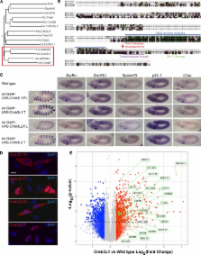
          CrebA up-regulates expression of both the general protein machinery required in all cells for secretion and genes encoding cell type–specific secreted components.

          Abstract

          Secretion occurs in all cells, with relatively low levels in most cells and extremely high levels in specialized secretory cells, such as those of the pancreas, salivary, and mammary glands. How secretory capacity is selectively up-regulated in specialized secretory cells is unknown. Here, we find that the CrebA/Creb3-like family of bZip transcription factors functions to up-regulate expression of both the general protein machinery required in all cells for secretion and of cell type–specific secreted proteins. Drosophila CrebA directly binds the enhancers of secretory pathway genes and is both necessary and sufficient to activate expression of every secretory pathway component gene examined thus far. Microarray profiling reveals that CrebA also up-regulates expression of genes encoding cell type–specific secreted components. Finally, we found that the human CrebA orthologues, Creb3L1 and Creb3L2, have the ability to up-regulate the secretory pathway in nonsecretory cell types.

          Related collections

          Most cited references35

          • Record: found
          • Abstract: found
          • Article: not found

          DAVID: Database for Annotation, Visualization, and Integrated Discovery.

          Functional annotation of differentially expressed genes is a necessary and critical step in the analysis of microarray data. The distributed nature of biological knowledge frequently requires researchers to navigate through numerous web-accessible databases gathering information one gene at a time. A more judicious approach is to provide query-based access to an integrated database that disseminates biologically rich information across large datasets and displays graphic summaries of functional information. Database for Annotation, Visualization, and Integrated Discovery (DAVID; http://www.david.niaid.nih.gov) addresses this need via four web-based analysis modules: 1) Annotation Tool - rapidly appends descriptive data from several public databases to lists of genes; 2) GoCharts - assigns genes to Gene Ontology functional categories based on user selected classifications and term specificity level; 3) KeggCharts - assigns genes to KEGG metabolic processes and enables users to view genes in the context of biochemical pathway maps; and 4) DomainCharts - groups genes according to PFAM conserved protein domains. Analysis results and graphical displays remain dynamically linked to primary data and external data repositories, thereby furnishing in-depth as well as broad-based data coverage. The functionality provided by DAVID accelerates the analysis of genome-scale datasets by facilitating the transition from data collection to biological meaning.
            Bookmark
            • Record: found
            • Abstract: found
            • Article: not found

            Endoplasmic reticulum stress activates cleavage of CREBH to induce a systemic inflammatory response.

            Regulated intramembrane proteolysis (RIP) of endoplasmic reticulum (ER) membrane-anchored transcription factors is known to maintain sterol homeostasis and to mediate the unfolded protein response (UPR). Here, we identified CREBH as a RIP-regulated liver-specific transcription factor that is cleaved upon ER stress and required to activate expression of acute phase response (APR) genes. Proinflammatory cytokines increase expression of ER membrane-anchored CREBH. In response to ER stress, CREBH is cleaved by site-1 and site-2 proteases to liberate an amino-terminal fragment that transits to the nucleus to activate transcription of the genes encoding serum amyloid P-component (SAP) and C-reactive protein (CRP). Proinflammatory cytokines and lipopolysaccharide activate the UPR and induce cleavage of CREBH in the liver in vivo. Together, our studies delineate a molecular mechanism for activation of an ER-localized transcription factor, CREBH, and reveal an unprecedented link by which ER stress initiates an acute inflammatory response.
              Bookmark
              • Record: found
              • Abstract: found
              • Article: not found

              ATF6 is a transcription factor specializing in the regulation of quality control proteins in the endoplasmic reticulum.

              Eukaryotic cells cope with endoplasmic reticulum (ER) stress by activating the unfolded protein response (UPR), a coordinated system of transcriptional and translational controls, which ensures the integrity of synthesized proteins. Mammalian cells express three UPR transducers in the ER, namely IRE1, PERK and ATF6. The IRE1 pathway, which is conserved from yeast to humans, mediates transcriptional induction of not only ER quality control proteins (molecular chaperones, folding enzymes and components of ER-associated degradation) but also proteins working at various stages of secretion. The PERK pathway, conserved in metazoan cells, is responsible for translational control and also participates in transcriptional control in mammals. ATF6 is an ER-membrane-bound transcription factor activated by ER stress-induced proteolysis which consists of two closely related factors, ATF6alpha and ATF6beta, in mammals. ATF6alpha but not ATF6beta plays an important role in transcriptional control. In this study, we performed a genome-wide search for ATF6alpha-target genes in mice. Only 30 of the 14,729 analyzable genes were identified as specific targets, of which 40% were ER quality control proteins, 20% were ER proteins, while the rest had miscellaneous functions. The negative effects of the absence of PERK on transcriptional induction of ER quality control proteins could be explained by its inhibitory effect on ATF6alpha activation. Further, proteins involved in transport from the ER are not regulated by ATF6alpha, and transport of folded cargo molecules from the ER was not affected by the absence of ATF6alpha. Based on these results, we propose that ATF6 is a transcription factor specialized in the regulation of ER quality control proteins.
                Bookmark

                Author and article information

                Journal
                J Cell Biol
                J. Cell Biol
                jcb
                The Journal of Cell Biology
                The Rockefeller University Press
                0021-9525
                1540-8140
                1 November 2010
                : 191
                : 3
                : 479-492
                Affiliations
                Department of Cell Biology, The Johns Hopkins University, School of Medicine, Baltimore, MD 21205
                Author notes
                Correspondence to Deborah J. Andrew: dandrew@ 123456jhmi.edu
                Article
                201004062
                10.1083/jcb.201004062
                3003312
                21041443
                dd727f29-c44f-4a93-a900-10d4f5e7a9f6
                © 2010 Fox et al.

                This article is distributed under the terms of an Attribution–Noncommercial–Share Alike–No Mirror Sites license for the first six months after the publication date (see http://www.rupress.org/terms). After six months it is available under a Creative Commons License (Attribution–Noncommercial–Share Alike 3.0 Unported license, as described at http://creativecommons.org/licenses/by-nc-sa/3.0/).

                History
                : 12 April 2010
                : 5 October 2010
                Categories
                Research Articles
                Article

                Cell biology
                Cell biology

                Comments

                Comment on this article