24
views
0
recommends
+1 Recommend
0 collections
    0
    shares
      • Record: found
      • Abstract: found
      • Article: not found

      Connexins in Cancer: Bridging the Gap to the Clinic

      research-article

      Read this article at

      ScienceOpenPublisherPMC
      Bookmark
          There is no author summary for this article yet. Authors can add summaries to their articles on ScienceOpen to make them more accessible to a non-specialist audience.

          Abstract

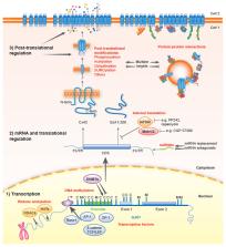
          Gap junctions comprise arrays of intercellular channels formed by connexin proteins and provide for direct communication between adjacent cells. This type of intercellular communication permits coordination of cellular activity and plays key roles in the control of cell growth and differentiation and in the maintenance of tissue homeostasis. After more than 50 years deciphering the links among connexins, gap junctions and cancer, researchers are now beginning to translate this knowledge to the clinic. The emergence of new strategies for connexin targeting, combined with improved understanding of the molecular bases underlying the dysregulation of connexins during cancer development, offer novel opportunities for clinical applications. However, different connexin isoforms have diverse channel-dependent and -independent functions that are tissue- and stage-specific. This can elicit both pro- and anti-tumorigenic effects that engender significant challenges in the path towards personalised medicine. Here, we review the current understanding of the role of connexins and gap junctions in cancer, with a particular focus on recent progress made in determining their prognostic and therapeutic potential.

          Related collections

          Most cited references212

          • Record: found
          • Abstract: found
          • Article: not found

          Carcinoma-astrocyte gap junctions promote brain metastasis by cGAMP transfer

          SUMMARY Brain metastasis represents a substantial source of morbidity and mortality in various cancers, and is characterized by high resistance to chemotherapy. Here we define the role of the most abundant cell type in the brain, the astrocyte, in promoting brain metastasis. Breast and lung cancer cells express protocadherin 7 (PCDH7) to favor the assembly of carcinoma-astrocyte gap junctions composed of connexin 43 (Cx43). Once engaged with the astrocyte gap-junctional network, brain metastatic cancer cells employ these channels to transfer the second messenger cGAMP to astrocytes, activating the STING pathway and production of inflammatory cytokines IFNα and TNFα. As paracrine signals, these factors activate the STAT1 and NF-κB pathways in brain metastatic cells, which support tumour growth and chemoresistance. The orally bioavailable modulators of gap junctions meclofenamate and tonabersat break this paracrine loop, and we provide proof-of-principle for the applicability of this therapeutic strategy to treat established brain metastasis.
            Bookmark
            • Record: found
            • Abstract: found
            • Article: not found

            Brain tumour cells interconnect to a functional and resistant network.

            Astrocytic brain tumours, including glioblastomas, are incurable neoplasms characterized by diffusely infiltrative growth. Here we show that many tumour cells in astrocytomas extend ultra-long membrane protrusions, and use these distinct tumour microtubes as routes for brain invasion, proliferation, and to interconnect over long distances. The resulting network allows multicellular communication through microtube-associated gap junctions. When damage to the network occurred, tumour microtubes were used for repair. Moreover, the microtube-connected astrocytoma cells, but not those remaining unconnected throughout tumour progression, were protected from cell death inflicted by radiotherapy. The neuronal growth-associated protein 43 was important for microtube formation and function, and drove microtube-dependent tumour cell invasion, proliferation, interconnection, and radioresistance. Oligodendroglial brain tumours were deficient in this mechanism. In summary, astrocytomas can develop functional multicellular network structures. Disconnection of astrocytoma cells by targeting their tumour microtubes emerges as a new principle to reduce the treatment resistance of this disease.
              Bookmark
              • Record: found
              • Abstract: found
              • Article: found
              Is Open Access

              Exosomes in Cancer Diagnostics

              Exosomes are endosome derived extracellular vesicles of 30–120 nm size ranges. Exosomes have been identified as mediators of cell-to-cell communication by transferring bioactive molecules such as nucleic acids, proteins and lipids into recipient cells. While exosomes are secreted by multiple cell types, cancer derived exosomes not only influence the invasive potentials of proximally located cells, but also affect distantly located tissues. Based on their ability to alter tumor microenvironment by regulating immunity, angiogenesis and metastasis, there has been growing interest in defining the clinical relevance of exosomes in cancers. In particular, exosomes are valuable sources for biomarkers due to selective cargo loading and resemblance to their parental cells. In this review, we summarize the recent findings to utilize exosomes as cancer biomarkers for early detection, diagnosis and therapy selection.
                Bookmark

                Author and article information

                Journal
                8711562
                Oncogene
                Oncogene
                Oncogene
                0950-9232
                1476-5594
                7 February 2019
                27 February 2019
                June 2019
                27 August 2019
                : 38
                : 23
                : 4429-4451
                Affiliations
                [1 ]Translational Molecular Pathology, Vall d'Hebron Institute of Research (VHIR), Autonomous University of Barcelona, CIBERONC, Barcelona, Spain
                [2 ]Department of Molecular Oncology, Institute for Cancer Research, Oslo University Hospital and K.G. Jebsen Colorectal Cancer Research Centre, Oslo University Hospital, Oslo, Norway
                [3 ]MRC-University of Glasgow Centre for Virus Research, Institute of Infection, Immunity and Inflammation, College of Medical Veterinary and Life Sciences, University of Glasgow, Glasgow, UK
                [4 ]Walter Brendel Centre of Experimental Medicine, Ludwig-Maximilians-Universität München and Munich University Hospital, München, Germany
                [5 ]CellCOM Research Group, Instituto de Investigación Biomédica de A Coruña (INIBIC), Servizo Galego de Saúde (SERGAS), University of A Coruña, A Coruña, Spain
                [6 ]STIM Laboratory, Faculté des Sciences Fondamentales et Appliquées, Université de Poitiers, Poitiers, France
                [7 ]Walter Brendel Centre of Experimental Medicine, Ludwig-Maximilians-Universität München and Munich University Hospital, München, Germany
                [8 ]Departamento de Bioquímica y Biología Molecular, Instituto de Neurociencias de Castilla y León (INCYL), Universidad de Salamanca, Salamanca, Spain
                Author notes
                Article
                EMS81526
                10.1038/s41388-019-0741-6
                6555763
                30814684
                2e82e535-e80f-49c3-93e9-6c697ce9ffc2

                Users may view, print, copy, and download text and data-mine the content in such documents, for the purposes of academic research, subject always to the full Conditions of use: http://www.nature.com/authors/editorial_policies/license.html#terms

                History
                Categories
                Article

                Oncology & Radiotherapy
                Oncology & Radiotherapy

                Comments

                Comment on this article