7
views
0
recommends
+1 Recommend
0 collections
    0
    shares
      • Record: found
      • Abstract: found
      • Article: found
      Is Open Access

      Mouse closed head traumatic brain injury replicates the histological tau pathology pattern of human disease: characterization of a novel model and systematic review of the literature

      research-article

      Read this article at

      Bookmark
          There is no author summary for this article yet. Authors can add summaries to their articles on ScienceOpen to make them more accessible to a non-specialist audience.

          Abstract

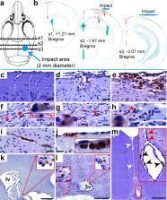
          Traumatic brain injury (TBI) constitutes one of the strongest environmental risk factors for several progressive neurodegenerative disorders of cognitive impairment and dementia that are characterized by the pathological accumulation of hyperphosphorylated tau (p-Tau). It has been questioned whether mouse closed-head TBI models can replicate human TBI-associated tauopathy. We conducted longitudinal histopathological characterization of a mouse closed head TBI model, with a focus on pathological features reported in human TBI-associated tauopathy. Male C57BL/6 J mice were subjected to once daily TBI for 5 consecutive days using a weight drop paradigm. Histological analyses (AT8, TDP-43, pTDP-43, NeuN, GFAP, Iba-1, MBP, SMI-312, Prussian blue, IgG, βAPP, alpha-synuclein) were conducted at 1 week, 4 weeks, and 24 weeks after rTBI and compared to sham operated controls. We conducted a systematic review of the literature for mouse models of closed-head injury focusing on studies referencing tau protein assessment. At 1-week post rTBI, p-Tau accumulation was restricted to the corpus callosum and perivascular spaces adjacent to the superior longitudinal fissure. Progressive p-Tau accumulation was observed in the superficial layers of the cerebral cortex, as well as in mammillary bodies and cortical perivascular, subpial, and periventricular locations at 4 to 24 weeks after rTBI. Associated cortical histopathologies included microvascular injury, neuroaxonal rarefaction, astroglial and microglial activation, and cytoplasmatic localization of TDP-43 and pTDP-43. In our systematic review, less than 1% of mouse studies (25/3756) reported p-Tau using immunostaining, of which only 3 (0.08%) reported perivascular p-Tau, which is considered a defining feature of chronic traumatic encephalopathy. Commonly reported associated pathologies included neuronal loss (23%), axonal loss (43%), microglial activation and astrogliosis (50%, each), and beta amyloid deposition (29%). Our novel model, supported by systematic review of the literature, indicates progressive tau pathology after closed head murine TBI, highlighting the suitability of mouse models to replicate pertinent human histopathology.

          Supplementary Information

          The online version contains supplementary material available at 10.1186/s40478-021-01220-8.

          Related collections

          Most cited references75

          • Record: found
          • Abstract: found
          • Article: not found

          NIH Image to ImageJ: 25 years of image analysis

          For the past twenty five years the NIH family of imaging software, NIH Image and ImageJ have been pioneers as open tools for scientific image analysis. We discuss the origins, challenges and solutions of these two programs, and how their history can serve to advise and inform other software projects.
            Bookmark
            • Record: found
            • Abstract: found
            • Article: not found

            Synapse loss and microglial activation precede tangles in a P301S tauopathy mouse model.

            Filamentous tau inclusions are hallmarks of Alzheimer's disease (AD) and related tauopathies, but earlier pathologies may herald disease onset. To investigate this, we studied wild-type and P301S mutant human tau transgenic (Tg) mice. Filamentous tau lesions developed in P301S Tg mice at 6 months of age, and progressively accumulated in association with striking neuron loss as well as hippocampal and entorhinal cortical atrophy by 9-12 months of age. Remarkably, hippocampal synapse loss and impaired synaptic function were detected in 3 month old P301S Tg mice before fibrillary tau tangles emerged. Prominent microglial activation also preceded tangle formation. Importantly, immunosuppression of young P301S Tg mice with FK506 attenuated tau pathology and increased lifespan, thereby linking neuroinflammation to early progression of tauopathies. Thus, hippocampal synaptic pathology and microgliosis may be the earliest manifestations of neurodegenerative tauopathies, and abrogation of tau-induced microglial activation could retard progression of these disorders.
              Bookmark
              • Record: found
              • Abstract: found
              • Article: not found

              Clinicopathological Evaluation of Chronic Traumatic Encephalopathy in Players of American Football.

              Players of American football may be at increased risk of long-term neurological conditions, particularly chronic traumatic encephalopathy (CTE).
                Bookmark

                Author and article information

                Contributors
                nils.henninger@umassmed.edu
                Journal
                Acta Neuropathol Commun
                Acta Neuropathol Commun
                Acta Neuropathologica Communications
                BioMed Central (London )
                2051-5960
                29 June 2021
                29 June 2021
                2021
                : 9
                : 118
                Affiliations
                [1 ]GRID grid.168645.8, ISNI 0000 0001 0742 0364, Department of Neurology, Medical School, , University of Massachusetts, ; 55 Lake Ave, Worcester, USA
                [2 ]GRID grid.168645.8, ISNI 0000 0001 0742 0364, Department of Pathology, Medical School, , University of Massachusetts, ; 55 Lake Ave, Worcester, USA
                [3 ]GRID grid.266683.f, ISNI 0000 0001 2184 9220, Department of Biology, , University of Massachusetts Amherst, ; Amherst, MA 01003 USA
                [4 ]GRID grid.168645.8, ISNI 0000 0001 0742 0364, Department of Psychiatry, Medical School, , University of Massachusetts, ; 55 Lake Ave, Worcester, USA
                Author information
                http://orcid.org/0000-0002-3883-5623
                Article
                1220
                10.1186/s40478-021-01220-8
                8243463
                34187585
                ddda207b-e326-4e39-804d-ca5cadb39af3
                © The Author(s) 2021

                Open AccessThis article is licensed under a Creative Commons Attribution 4.0 International License, which permits use, sharing, adaptation, distribution and reproduction in any medium or format, as long as you give appropriate credit to the original author(s) and the source, provide a link to the Creative Commons licence, and indicate if changes were made. The images or other third party material in this article are included in the article's Creative Commons licence, unless indicated otherwise in a credit line to the material. If material is not included in the article's Creative Commons licence and your intended use is not permitted by statutory regulation or exceeds the permitted use, you will need to obtain permission directly from the copyright holder. To view a copy of this licence, visit http://creativecommons.org/licenses/by/4.0/. The Creative Commons Public Domain Dedication waiver ( http://creativecommons.org/publicdomain/zero/1.0/) applies to the data made available in this article, unless otherwise stated in a credit line to the data.

                History
                : 11 May 2021
                : 18 June 2021
                Funding
                Funded by: FundRef http://dx.doi.org/10.13039/100000065, National Institute of Neurological Disorders and Stroke;
                Award ID: K08NS091499
                Award Recipient :
                Categories
                Research
                Custom metadata
                © The Author(s) 2021

                animal model,chronic traumatic encephalopathy,concussion,systematic review,tauopathy,traumatic brain injury

                Comments

                Comment on this article