58
views
0
recommends
+1 Recommend
0 collections
    0
    shares
      • Record: found
      • Abstract: found
      • Article: found
      Is Open Access

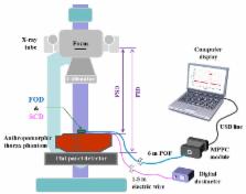
      Measurement of Entrance Surface Dose on an Anthropomorphic Thorax Phantom Using a Miniature Fiber-Optic Dosimeter

      research-article

      Read this article at

      Bookmark
          There is no author summary for this article yet. Authors can add summaries to their articles on ScienceOpen to make them more accessible to a non-specialist audience.

          Abstract

          A miniature fiber-optic dosimeter (FOD) system was fabricated using a plastic scintillating fiber, a plastic optical fiber, and a multi-pixel photon counter to measure real-time entrance surface dose (ESD) during radiation diagnosis. Under varying exposure parameters of a digital radiography (DR) system, we measured the scintillating light related to the ESD using the sensing probe of the FOD, which was placed at the center of the beam field on an anthropomorphic thorax phantom. Also, we obtained DR images using a flat panel detector of the DR system to evaluate the effects of the dosimeter on image artifacts during posteroanterior (PA) chest radiography. From the experimental results, the scintillation output signals of the FOD were similar to the ESDs including backscatter simultaneously obtained using a semiconductor dosimeter. We demonstrated that the proposed miniature FOD can be used to measure real-time ESDs with minimization of DR image artifacts in the X-ray energy range of diagnostic radiology.

          Related collections

          Most cited references22

          • Record: found
          • Abstract: found
          • Article: not found

          Miniature scintillating detector for small field radiation therapy.

          In planning stereotactic radiosurgery treatments, depth dose curves, profiles, and dose rate of treatment beams are difficult to obtain with conventional detectors because of loss of lateral electronic equilibrium and volume averaging. A scintillating detector with high spatial resolution and good reliability has been developed to overcome this problem. The miniature dosimeter consists of two identical radiation-resistant 10 m long silica optical fibers, each connected to an independent silicon photodiode. A small cylindrical polystyrene scintillator (3.9 mm3) is optically glued to the detection fiber. The light seen by the photodiode connected to this fiber arises from fluorescence of the scintillator and from the Cerenkov effect produced in silica. The reference signal produced by the fiber without scintillator is used to subtract the Cerenkov light contribution from the raw detector response. The sensitive volume of the scintillating detector is nearly water-equivalent and thus minimizes dose distribution perturbation in water. The miniature dosimeter has a spatial resolution comparable to the film-densitometer system. Profiles of 1 cm diam, 6 MV photon beam measured with both systems show very similar shapes. Furthermore, the use of photodiodes instead of photomultiplier tubes gives a better stability response and offers the possibility to perform absolute dosimetry.
            Bookmark
            • Record: found
            • Abstract: found
            • Article: not found

            Plastic scintillator response to low-energy photons.

            The plastic scintillator (PS) is a promising dosimeter for brachytherapy and other low-energy photon applications because of its high sensitivity and approximate tissue equivalence. As part of our project to develop a new PS material which maximizes sensitivity and radiological equivalence to water, we have measured the response, epsilon (light output/unit air kerma), of PS to low-energy bremsstrahlung (20 to 57 keV average energies) x-rays as well as photons emitted by 99mTc, 192Ir, and 137Cs sources, all of which were calibrated in terms of air kerma. The PS systems studied were a standard commercial PS, BC400 (Bicron Corporation, Newbury, OH), and our new sensitive and quench-resistant scintillator (polyvinyltoluene base and binary dye system) with and without 4% Cl loading intended to match the effective atomic number of water. For low-energy x-rays, epsilon was 20-57% relative to epsilon for 192Ir photons. Chlorine loading clearly reduced the energy dependence of epsilon, which ranged from 46% to 85% relative to 192Ir. However, even after using Monte Carlo photon-transport simulation to correct for the non-air equivalence of the PS, inherent dosimetric sensitivity still varied by 30% over the 20-400 keV energy range. Our work, one of the few measurements of PS response to low-energy photons, appears to confirm Birks' 1955 finding that ionization quenching reduces sensitivity to electrons below 125 keV. However, our results cannot be explained by Birks' widely used unimolecular quenching model.
              Bookmark
              • Record: found
              • Abstract: found
              • Article: not found

              Validating plastic scintillation detectors for photon dosimetry in the radiologic energy range.

              Photon dosimetry in the kilovolt (kV) energy range represents a major challenge for diagnostic and interventional radiology and superficial therapy. Plastic scintillation detectors (PSDs) are potentially good candidates for this task. This study proposes a simple way to obtain accurate correction factors to compensate for the response of PSDs to photon energies between 80 and 150 kVp. The performance of PSDs is also investigated to determine their potential usefulness in the diagnostic energy range. A 1-mm-diameter, 10-mm-long PSD was irradiated by a Therapax SXT 150 unit using five different beam qualities made of tube potentials ranging from 80 to 150 kVp and filtration thickness ranging from 0.8 to 0.2 mmAl + 1.0 mmCu. The light emitted by the detector was collected using an 8-m-long optical fiber and a polychromatic photodiode, which converted the scintillation photons to an electrical current. The PSD response was compared with the reference free air dose rate measured with a calibrated Farmer NE2571 ionization chamber. PSD measurements were corrected using spectra-weighted corrections, accounting for mass energy-absorption coefficient differences between the sensitive volumes of the ionization chamber and the PSD, as suggested by large cavity theory (LCT). Beam spectra were obtained from x-ray simulation software and validated experimentally using a CdTe spectrometer. Correction factors were also obtained using Monte Carlo (MC) simulations. Percent depth dose (PDD) measurements were compensated for beam hardening using the LCT correction method. These PDD measurements were compared with uncorrected PSD data, PDD measurements obtained using Gafchromic films, Monte Carlo simulations, and previous data. For each beam quality used, the authors observed an increase of the energy response with effective energy when no correction was applied to the PSD response. Using the LCT correction, the PSD response was almost energy independent, with a residual 2.1% coefficient of variation (COV) over the 80-150-kVp energy range. Monte Carlo corrections reduced the COV to 1.4% over this energy range. All PDD measurements were in good agreement with one another except for the uncorrected PSD data, in which an over-response was observed with depth (13% at 10 cm with a 100 kVp beam), showing that beam hardening had a non-negligible effect on the PSD response. A correction based on LCT compensated very well for this effect, reducing the over-response to 3%. In the diagnostic energy range, PSDs show high-energy dependence, which can be corrected using spectra-weighted mass energy-absorption coefficients, showing no considerable sign of quenching between these energies. Correction factors obtained by Monte Carlo simulations confirm that the approximations made by LCT corrections are valid. Thus, PSDs could be useful for real-time dosimetry in radiology applications.
                Bookmark

                Author and article information

                Journal
                Sensors (Basel)
                Sensors (Basel)
                Sensors (Basel, Switzerland)
                Molecular Diversity Preservation International (MDPI)
                1424-8220
                April 2014
                01 April 2014
                : 14
                : 4
                : 6305-6316
                Affiliations
                [1 ] School of Biomedical Engineering, College of Biomedical & Health Science, Research Institute of Biomedical Engineering, Konkuk University, 268 Chungwon-daero, Chungju-si, Chungcheongbuk-do, 380-701, Korea; E-Mails: wonzip@ 123456kku.ac.kr (W.J.Y.); shshin9431@ 123456gmail.com (S.H.S); jdy603@ 123456naver.com (D.J); zzang811@ 123456gmail.com (S.H.); saucony9116@ 123456msn.com (H.I.S.); chokomilkys@ 123456gmail.com (S.G.K.); kko988@ 123456kku.ac.kr (K.W.J.)
                [2 ] Department of Organic Materials & Fiber Engineering, College of Engineering, Soongsil University, 369 Sangdo-ro, Dongjak-gu, Seoul-si, 156-743, Korea; E-Mail: scho@ 123456ssu.ac.kr
                [3 ] Research & Development Center, JPI Healthcare, Osongsaengmyeong 1-ro, Osong-eup, Cheongwon-gun, Chungcheongbuk-do, 363-951, Korea; E-Mail: younws@ 123456JPI.co.kr
                Author notes

                Author Contributions Wook Jae Yoo made substantial contributions in design of the sensor, all experiments, data analysis, and manuscript preparation. Bongsoo Lee made significant contributions in experimental design, data analysis and manuscript preparation. Sang Hun Shin, Dayeong Jeon, Seunghan Hong, Hyeok In Sim, and Seon Geun Kim made contributions in fabrication of the sensor, experimental setup and data acquisition. Kyoung Won Jang and Seunghyun Cho made contributions in data analysis and sensor characterization. Won Sik Youn provided digital radiography system and made contributions in experimental design and data acquisition.

                [* ] Author to whom correspondence should be addressed; E-Mail: bslee@ 123456kku.ac.kr ; Tel.: +82-43-840-3755; Fax: +82-43-851-0620.
                Article
                sensors-14-06305
                10.3390/s140406305
                4029715
                24694678
                cd9170b2-9272-40f3-bf1b-e0a2c0501e3c
                © 2014 by the authors; licensee MDPI, Basel, Switzerland.

                This article is an open access article distributed under the terms and conditions of the Creative Commons Attribution license ( http://creativecommons.org/licenses/by/3.0/).

                History
                : 24 December 2013
                : 26 March 2014
                : 27 March 2014
                Categories
                Article

                Biomedical engineering
                fiber-optic dosimeter,entrance surface dose,diagnostic radiology,scintillating light,anthropomorphic thorax phantom

                Comments

                Comment on this article