62
views
0
recommends
+1 Recommend
0 collections
    0
    shares
      • Record: found
      • Abstract: found
      • Article: found
      Is Open Access

      Deep learning for lung cancer prognostication: A retrospective multi-cohort radiomics study

      research-article

      Read this article at

      Bookmark
          There is no author summary for this article yet. Authors can add summaries to their articles on ScienceOpen to make them more accessible to a non-specialist audience.

          Abstract

          Background

          Non-small-cell lung cancer (NSCLC) patients often demonstrate varying clinical courses and outcomes, even within the same tumor stage. This study explores deep learning applications in medical imaging allowing for the automated quantification of radiographic characteristics and potentially improving patient stratification.

          Methods and findings

          We performed an integrative analysis on 7 independent datasets across 5 institutions totaling 1,194 NSCLC patients (age median = 68.3 years [range 32.5–93.3], survival median = 1.7 years [range 0.0–11.7]). Using external validation in computed tomography (CT) data, we identified prognostic signatures using a 3D convolutional neural network (CNN) for patients treated with radiotherapy ( n = 771, age median = 68.0 years [range 32.5–93.3], survival median = 1.3 years [range 0.0–11.7]). We then employed a transfer learning approach to achieve the same for surgery patients ( n = 391, age median = 69.1 years [range 37.2–88.0], survival median = 3.1 years [range 0.0–8.8]). We found that the CNN predictions were significantly associated with 2-year overall survival from the start of respective treatment for radiotherapy (area under the receiver operating characteristic curve [AUC] = 0.70 [95% CI 0.63–0.78], p < 0.001) and surgery (AUC = 0.71 [95% CI 0.60–0.82], p < 0.001) patients. The CNN was also able to significantly stratify patients into low and high mortality risk groups in both the radiotherapy ( p < 0.001) and surgery ( p = 0.03) datasets. Additionally, the CNN was found to significantly outperform random forest models built on clinical parameters—including age, sex, and tumor node metastasis stage—as well as demonstrate high robustness against test–retest (intraclass correlation coefficient = 0.91) and inter-reader (Spearman’s rank-order correlation = 0.88) variations. To gain a better understanding of the characteristics captured by the CNN, we identified regions with the most contribution towards predictions and highlighted the importance of tumor-surrounding tissue in patient stratification. We also present preliminary findings on the biological basis of the captured phenotypes as being linked to cell cycle and transcriptional processes. Limitations include the retrospective nature of this study as well as the opaque black box nature of deep learning networks.

          Conclusions

          Our results provide evidence that deep learning networks may be used for mortality risk stratification based on standard-of-care CT images from NSCLC patients. This evidence motivates future research into better deciphering the clinical and biological basis of deep learning networks as well as validation in prospective data.

          Abstract

          Hugo Aerts and colleagues evaluate the ability of deep learning networks to extract relevant features from computed tomography lung cancer images and stratify patients into low and high mortality risk groups.

          Author summary

          Why was this study done?
          • Cancer is one of the leading causes of death worldwide, with lung cancer being the second most commonly diagnosed cancer in both men and women in the US.

          • Prognosis in lung cancer patients is primarily determined through tumor staging, which in turn is based on a relatively coarse and discrete stratification.

          • Radiographic medical images offer patient- and tumor-specific information that could be used to complement clinical prognostic evaluation efforts.

          • Recent advances in radiomics through applications of artificial intelligence, computer vision, and deep learning allow for the extraction and mining of numerous quantitative features from radiographic images.

          What did the researchers do and find?
          • We designed an analysis setup comprising 7 independent datasets across 5 institutions totaling 1,194 patients with non-small-cell lung cancer imaged with computed tomography and treated with either radiotherapy or surgery.

          • We evaluated the prognostic signatures of quantitative imaging features extracted through deep learning networks, and assessed their ability to stratify patients into low and high mortality risk groups as per a 2-year overall survival cutoff.

          • In patients treated with surgery, deep learning networks significantly outperformed models based on predefined tumor features as well as tumor volume and maximum diameter.

          • In addition to highlighting image regions with prognostic influence, we evaluated the deep learning features for robustness against physiological imaging artifacts and input variability, as well as correlated them with molecular information through gene expression data.

          What do these findings mean?
          • We found that deep learning features significantly outperform existing prognostication methods in surgery patients, hinting at their utility in patient stratification and potentially sparing low mortality risk groups from adjuvant chemotherapy.

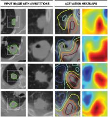
          • We demonstrated that areas within and beyond the tumor—especially the tumor–stroma interfaces—had the largest contributions to the prognostic signature, highlighting the importance of tumor-surrounding tissue in patient stratification.

          • Preliminary genomic associations in this study suggest correlations between the deep learning feature representations and cell cycle and transcriptional processes.

          • Despite their obscure inner workings and lack of a strong theoretical backing, deep learning networks demonstrate a prognostic signal and robustness against specific noise artifacts. This finding motivates further prospective studies validating their utility in patient stratification and the development of personalized cancer treatment plans.

          Related collections

          Most cited references39

          • Record: found
          • Abstract: not found
          • Article: not found

          Controlling the False Discovery Rate: A Practical and Powerful Approach to Multiple Testing

            Bookmark
            • Record: found
            • Abstract: found
            • Article: found
            Is Open Access

            Machine Learning methods for Quantitative Radiomic Biomarkers

            Radiomics extracts and mines large number of medical imaging features quantifying tumor phenotypic characteristics. Highly accurate and reliable machine-learning approaches can drive the success of radiomic applications in clinical care. In this radiomic study, fourteen feature selection methods and twelve classification methods were examined in terms of their performance and stability for predicting overall survival. A total of 440 radiomic features were extracted from pre-treatment computed tomography (CT) images of 464 lung cancer patients. To ensure the unbiased evaluation of different machine-learning methods, publicly available implementations along with reported parameter configurations were used. Furthermore, we used two independent radiomic cohorts for training (n = 310 patients) and validation (n = 154 patients). We identified that Wilcoxon test based feature selection method WLCX (stability = 0.84 ± 0.05, AUC = 0.65 ± 0.02) and a classification method random forest RF (RSD = 3.52%, AUC = 0.66 ± 0.03) had highest prognostic performance with high stability against data perturbation. Our variability analysis indicated that the choice of classification method is the most dominant source of performance variation (34.21% of total variance). Identification of optimal machine-learning methods for radiomic applications is a crucial step towards stable and clinically relevant radiomic biomarkers, providing a non-invasive way of quantifying and monitoring tumor-phenotypic characteristics in clinical practice.
              Bookmark
              • Record: found
              • Abstract: found
              • Article: found
              Is Open Access

              A Deep Learning-Based Radiomics Model for Prediction of Survival in Glioblastoma Multiforme

              Traditional radiomics models mainly rely on explicitly-designed handcrafted features from medical images. This paper aimed to investigate if deep features extracted via transfer learning can generate radiomics signatures for prediction of overall survival (OS) in patients with Glioblastoma Multiforme (GBM). This study comprised a discovery data set of 75 patients and an independent validation data set of 37 patients. A total of 1403 handcrafted features and 98304 deep features were extracted from preoperative multi-modality MR images. After feature selection, a six-deep-feature signature was constructed by using the least absolute shrinkage and selection operator (LASSO) Cox regression model. A radiomics nomogram was further presented by combining the signature and clinical risk factors such as age and Karnofsky Performance Score. Compared with traditional risk factors, the proposed signature achieved better performance for prediction of OS (C-index = 0.710, 95% CI: 0.588, 0.932) and significant stratification of patients into prognostically distinct groups (P < 0.001, HR = 5.128, 95% CI: 2.029, 12.960). The combined model achieved improved predictive performance (C-index = 0.739). Our study demonstrates that transfer learning-based deep features are able to generate prognostic imaging signature for OS prediction and patient stratification for GBM, indicating the potential of deep imaging feature-based biomarker in preoperative care of GBM patients.
                Bookmark

                Author and article information

                Contributors
                Role: ConceptualizationRole: Data curationRole: Formal analysisRole: MethodologyRole: SoftwareRole: ValidationRole: VisualizationRole: Writing – original draftRole: Writing – review & editing
                Role: ConceptualizationRole: Formal analysisRole: MethodologyRole: ValidationRole: Writing – review & editing
                Role: Data curationRole: MethodologyRole: ValidationRole: Writing – review & editing
                Role: Formal analysisRole: SoftwareRole: Writing – review & editing
                Role: Formal analysisRole: SoftwareRole: Writing – review & editing
                Role: Formal analysisRole: SoftwareRole: Writing – review & editing
                Role: Data curationRole: Writing – review & editing
                Role: Data curationRole: MethodologyRole: Writing – review & editing
                Role: Data curationRole: InvestigationRole: MethodologyRole: Project administrationRole: SupervisionRole: Writing – review & editing
                Role: ConceptualizationRole: Funding acquisitionRole: MethodologyRole: Project administrationRole: SupervisionRole: Writing – review & editing
                Role: Academic Editor
                Journal
                PLoS Med
                PLoS Med
                plos
                plosmed
                PLoS Medicine
                Public Library of Science (San Francisco, CA USA )
                1549-1277
                1549-1676
                30 November 2018
                November 2018
                : 15
                : 11
                : e1002711
                Affiliations
                [1 ] Department of Radiation Oncology, Dana-Farber Cancer Institute, Brigham and Women’s Hospital, Harvard Medical School, Boston, Massachusetts, United States of America
                [2 ] Department of Radiation Oncology, Radboud University Medical Center, Nijmegen, The Netherlands
                [3 ] Department of Cancer Physiology, H. Lee Moffitt Cancer Center and Research Institute, Tampa, Florida, United States of America
                [4 ] Department of Radiology, Brigham and Women’s Hospital, Harvard Medical School, Boston, Massachusetts, United States of America
                University of California San Francisco, UNITED STATES
                Author notes

                I have read the journal’s policy and the authors of this manuscript have the following competing interests: RHM reports personal fees (Scientific Advisory Board) from AstraZeneca, personal fees from New RT (lecture honorarium), outside the submitted work. RJG is a consultant, shareholder and grantee of HealthMyne, Inc., and a consultant, shareholder and grantee of Helix Biopharma. Neither Helix nor HealthMyne have a financial stake in the results of this study. All other authors declare no conflict of interest.

                Author information
                http://orcid.org/0000-0002-1844-481X
                http://orcid.org/0000-0003-4918-6902
                http://orcid.org/0000-0001-5882-748X
                http://orcid.org/0000-0002-8888-7747
                http://orcid.org/0000-0002-2122-2003
                Article
                PMEDICINE-D-18-01161
                10.1371/journal.pmed.1002711
                6269088
                30500819
                c1a2965b-c8a2-4b40-a2bc-ab4f22585e14
                © 2018 Hosny et al

                This is an open access article distributed under the terms of the Creative Commons Attribution License, which permits unrestricted use, distribution, and reproduction in any medium, provided the original author and source are credited.

                History
                : 29 March 2018
                : 5 November 2018
                Page count
                Figures: 5, Tables: 0, Pages: 25
                Funding
                Funded by: funder-id http://dx.doi.org/10.13039/100000002, National Institutes of Health;
                Award ID: U24CA194354
                Award Recipient :
                Funded by: funder-id http://dx.doi.org/10.13039/100000002, National Institutes of Health;
                Award ID: U01CA190234
                Award Recipient :
                Authors acknowledge financial support from the National Institute of Health (NIH-USA U24CA194354, and NIH-USA U01CA190234); https://grants.nih.gov/funding/index.htm. The funders had no role in study design, data collection and analysis, decision to publish, or preparation of the manuscript.
                Categories
                Research Article
                Computer and Information Sciences
                Artificial Intelligence
                Machine Learning
                Deep Learning
                Medicine and Health Sciences
                Surgical and Invasive Medical Procedures
                Medicine and Health Sciences
                Oncology
                Cancer Treatment
                Radiation Therapy
                Medicine and Health Sciences
                Clinical Medicine
                Clinical Oncology
                Radiation Therapy
                Medicine and Health Sciences
                Oncology
                Clinical Oncology
                Radiation Therapy
                Medicine and Health Sciences
                Oncology
                Cancers and Neoplasms
                Lung and Intrathoracic Tumors
                Non-Small Cell Lung Cancer
                Medicine and Health Sciences
                Oncology
                Cancer Treatment
                Science Policy
                Science and Technology Workforce
                Careers in Research
                Engineers
                People and Places
                Population Groupings
                Professions
                Engineers
                Research and Analysis Methods
                Imaging Techniques
                Neuroimaging
                Computed Axial Tomography
                Biology and Life Sciences
                Neuroscience
                Neuroimaging
                Computed Axial Tomography
                Medicine and Health Sciences
                Diagnostic Medicine
                Diagnostic Radiology
                Tomography
                Computed Axial Tomography
                Research and Analysis Methods
                Imaging Techniques
                Diagnostic Radiology
                Tomography
                Computed Axial Tomography
                Medicine and Health Sciences
                Radiology and Imaging
                Diagnostic Radiology
                Tomography
                Computed Axial Tomography
                Medicine and Health Sciences
                Oncology
                Cancer Treatment
                Surgical Oncology
                Medicine and Health Sciences
                Clinical Medicine
                Clinical Oncology
                Surgical Oncology
                Medicine and Health Sciences
                Oncology
                Clinical Oncology
                Surgical Oncology
                Custom metadata
                The raw imaging data for the respective datasets can be accessed at: Maastro https://wiki.cancerimagingarchive.net/display/Public/NSCLC-Radiomics#38439daf994e4b5595a0d431342b4c33, MUMC https://wiki.cancerimagingarchive.net/display/Public/NSCLC-Radiomics-Genomics, and RIDER https://wiki.cancerimagingarchive.net/display/Public/RIDER+Collections. Tabular data containing 2 year survival information, convolutional neural network (CNN) predictions, and engineered features can be accessed at https://github.com/modelhub-ai/deep-prognosis/tree/master/data. CNN implementation can be accessed at https://github.com/modelhub-ai/deep-prognosis/tree/master/contrib_src/model. R analysis files containing AUC and p-value calculations for convolutional neural networks and other random forest models can be accessed at https://github.com/modelhub-ai/deep-prognosis/tree/master/R_files. Gene set enrichment analysis (GSEA) files can be accessed at https://github.com/modelhub-ai/deep-prognosis/tree/master/gsea. This analysis uses the GSEA desktop application developed by the Broad Institute and can be downloaded at http://software.broadinstitute.org/gsea/index.jsp.

                Medicine
                Medicine

                Comments

                Comment on this article