3
views
0
recommends
+1 Recommend
0 collections
    0
    shares
      • Record: found
      • Abstract: found
      • Article: found
      Is Open Access

      Superresolution microscopy localizes endogenous Dvl2 to Wnt signaling-responsive biomolecular condensates

      research-article

      Read this article at

      Bookmark
          There is no author summary for this article yet. Authors can add summaries to their articles on ScienceOpen to make them more accessible to a non-specialist audience.

          Significance

          Wnt signaling governs cell fate and tissue polarity across species. The Dishevelled proteins are central to Wnt signaling cascades. Wnt-mediated multiprotein complexes such as the “signalosome” and the “destruction complex” have been proposed to represent biomolecular condensates. These nonmembranous, specialized compartments have been suggested to form through liquid–liquid phase separation and ensure correctly proceeding physiological reactions. Although biomolecular condensates have increasingly been studied, key questions remain regarding, for example, their architecture and physiological regulation. Here, superresolution microscopy after endogenous labeling of Dishevelled-2 gives insights into protein functions and Wnt signaling at physiological levels. It reveals the distinct molecular architecture of endogenous Wnt condensates at single-molecule resolution and illustrates close interactions at the centrosome.

          Abstract

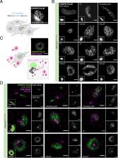
          During organismal development, homeostasis, and disease, Dishevelled (Dvl) proteins act as key signaling factors in beta-catenin–dependent and beta-catenin–independent Wnt pathways. While their importance for signal transmission has been genetically demonstrated in many organisms, our mechanistic understanding is still limited. Previous studies using overexpressed proteins showed Dvl localization to large, punctate-like cytoplasmic structures that are dependent on its DIX domain. To study Dvl’s role in Wnt signaling, we genome engineered an endogenously expressed Dvl2 protein tagged with an mEos3.2 fluorescent protein for superresolution imaging. First, we demonstrate the functionality and specificity of the fusion protein in beta-catenin–dependent and beta-catenin–independent signaling using multiple independent assays. We performed live-cell imaging of Dvl2 to analyze the dynamic formation of the supramolecular cytoplasmic Dvl2_mEos3.2 condensates. While overexpression of Dvl2_mEos3.2 mimics the previously reported formation of abundant large “puncta,” supramolecular condensate formation at physiological protein levels is only observed in a subset of cells with approximately one per cell. We show that, in these condensates, Dvl2 colocalizes with Wnt pathway components at gamma-tubulin and CEP164-positive centrosomal structures and that the localization of Dvl2 to these condensates is Wnt dependent. Single-molecule localization microscopy using photoactivated localization microscopy (PALM) of mEos3.2 in combination with DNA-PAINT demonstrates the organization and repetitive patterns of these condensates in a cell cycle–dependent manner. Our results indicate that the localization of Dvl2 in supramolecular condensates is coordinated dynamically and dependent on cell state and Wnt signaling levels. Our study highlights the formation of endogenous and physiologically regulated biomolecular condensates in the Wnt pathways at single-molecule resolution.

          Related collections

          Most cited references107

          • Record: found
          • Abstract: found
          • Article: not found

          Biomolecular condensates: organizers of cellular biochemistry

          In addition to membrane-bound organelles, eukaryotic cells feature various membraneless compartments, including the centrosome, the nucleolus and various granules. Many of these compartments form through liquid–liquid phase separation, and the principles, mechanisms and regulation of their assembly as well as their cellular functions are now beginning to emerge.
            Bookmark
            • Record: found
            • Abstract: found
            • Article: not found

            Wnt/β-catenin signaling and disease.

            The WNT signal transduction cascade controls myriad biological phenomena throughout development and adult life of all animals. In parallel, aberrant Wnt signaling underlies a wide range of pathologies in humans. In this Review, we provide an update of the core Wnt/β-catenin signaling pathway, discuss how its various components contribute to disease, and pose outstanding questions to be addressed in the future. Copyright © 2012 Elsevier Inc. All rights reserved.
              Bookmark
              • Record: found
              • Abstract: found
              • Article: not found

              Sub-diffraction-limit imaging by stochastic optical reconstruction microscopy (STORM).

              We have developed a high-resolution fluorescence microscopy method based on high-accuracy localization of photoswitchable fluorophores. In each imaging cycle, only a fraction of the fluorophores were turned on, allowing their positions to be determined with nanometer accuracy. The fluorophore positions obtained from a series of imaging cycles were used to reconstruct the overall image. We demonstrated an imaging resolution of 20 nm. This technique can, in principle, reach molecular-scale resolution.
                Bookmark

                Author and article information

                Journal
                Proc Natl Acad Sci U S A
                Proc Natl Acad Sci U S A
                pnas
                PNAS
                Proceedings of the National Academy of Sciences of the United States of America
                National Academy of Sciences
                0027-8424
                1091-6490
                22 July 2022
                26 July 2022
                22 July 2022
                : 119
                : 30
                : e2122476119
                Affiliations
                [1] aDivision of Signaling and Functional Genomics, German Cancer Research Center and Department of Cell and Molecular Biology, Heidelberg University and BioQuant , 69120 Heidelberg, Germany;
                [2] bDepartment of Hematology and Medical Oncology, University Medical Center Göttingen , 37075 Göttingen, Germany;
                [3] cDepartment of Medical Oncology, National Center for Tumor Diseases, University Hospital Heidelberg , 69120 Heidelberg, Germany;
                [4] dAdvanced Light Microscopy Facility, European Molecular Biology Laboratory, 69117 Heidelberg, Germany;
                [5] eNikon Imaging Center, BioQuant and Centre for Organismal Studies, Heidelberg University , 69120 Heidelberg, Germany
                Author notes
                3To whom correspondence may be addressed. Email: m.boutros@ 123456dkfz.de .

                Edited by Roel Nusse, Stanford University School of Medicine, Stanford, CA; received December 13, 2021; accepted April 15, 2022

                Author contributions: A.S., O.V., L.T., C.B., U.E., and M.B. designed research; A.S., O.V., F.R., P.G., C.S., T.M., M.S., M.L., and U.E. performed research; A.S., O.V., F.R., P.G., C.S., and U.E. contributed new reagents/analytic tools; A.S., O.V., F.R., P.G., C.S., M.L., and U.E. analyzed data; and A.S., D.K., and M.B. wrote the paper.

                1Present address: Science for Life Laboratory, Department of Women’s and Children’s Health, Karolinska Institutet, 17165 Solna, Sweden.

                2Present address: Department for Cardiovascular and Metabolic Diseases, Max Delbrück Center for Molecular Medicine in the Helmholtz Association, 13125 Berlin, Germany.

                Author information
                https://orcid.org/0000-0002-7455-5047
                https://orcid.org/0000-0002-0673-3278
                https://orcid.org/0000-0002-4148-262X
                https://orcid.org/0000-0002-5258-0321
                https://orcid.org/0000-0003-3604-7647
                https://orcid.org/0000-0003-1786-4916
                https://orcid.org/0000-0002-4510-9048
                https://orcid.org/0000-0002-6953-6845
                https://orcid.org/0000-0002-9458-817X
                Article
                202122476
                10.1073/pnas.2122476119
                9335300
                35867833
                bfa0bf6e-ab43-4037-99db-4571fe9d82c4
                Copyright © 2022 the Author(s). Published by PNAS.

                This open access article is distributed under Creative Commons Attribution-NonCommercial-NoDerivatives License 4.0 (CC BY-NC-ND).

                History
                : 15 April 2022
                Page count
                Pages: 12
                Funding
                Funded by: Deutsche Forschungsgemeinschaft (DFG) 501100001659
                Award ID: SFB1324
                Award Recipient : Antonia Schubert Award Recipient : Oksana Voloshanenko Award Recipient : Michael Boutros
                Categories
                409
                Biological Sciences
                Cell Biology

                wnt signaling,dishevelled,superresolution microscopy,biomolecular condensates,crispr

                Comments

                Comment on this article