2
views
0
recommends
+1 Recommend
1 collections
    0
    shares
      • Record: found
      • Abstract: found
      • Article: found
      Is Open Access

      SARS-CoV-2 requires acidic pH to infect cells

      research-article

      Read this article at

      Bookmark
          There is no author summary for this article yet. Authors can add summaries to their articles on ScienceOpen to make them more accessible to a non-specialist audience.

          Significance

          Infection by SARS-CoV-2 depends upon the large spike (S) protein decorating the virions and is responsible for receptor engagement and subsequent fusion of viral and cellular membranes allowing release of virion contents into the cell. Using new single-particle imaging tools to visualize and track the successive steps from virion attachment to fusion, combined with chemical and genetic perturbations of the cells, we provide direct evidence for the cellular uptake routes of productive infection in multiple cell types and their dependence on proteolysis of S by cell surface or endosomal proteases. We show that fusion and content release always require the acidic environment from endosomes, preceded by liberation of the S1 fragment which depends on angiotensin converting enzyme receptor engagement.

          Abstract

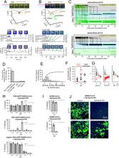
          Severe acute respiratory syndrome coronavirus 2 (SARS-CoV-2) cell entry starts with membrane attachment and ends with spike (S) protein–catalyzed membrane fusion depending on two cleavage steps, namely, one usually by furin in producing cells and the second by TMPRSS2 on target cells. Endosomal cathepsins can carry out both. Using real-time three-dimensional single-virion tracking, we show that fusion and genome penetration require virion exposure to an acidic milieu of pH 6.2 to 6.8, even when furin and TMPRSS2 cleavages have occurred. We detect the sequential steps of S1-fragment dissociation, fusion, and content release from the cell surface in TMPRRS2-overexpressing cells only when exposed to acidic pH. We define a key role of an acidic environment for successful infection, found in endosomal compartments and at the surface of TMPRSS2-expressing cells in the acidic milieu of the nasal cavity.

          Related collections

          Most cited references46

          • Record: found
          • Abstract: found
          • Article: not found

          SARS-CoV-2 Cell Entry Depends on ACE2 and TMPRSS2 and Is Blocked by a Clinically Proven Protease Inhibitor

          Summary The recent emergence of the novel, pathogenic SARS-coronavirus 2 (SARS-CoV-2) in China and its rapid national and international spread pose a global health emergency. Cell entry of coronaviruses depends on binding of the viral spike (S) proteins to cellular receptors and on S protein priming by host cell proteases. Unravelling which cellular factors are used by SARS-CoV-2 for entry might provide insights into viral transmission and reveal therapeutic targets. Here, we demonstrate that SARS-CoV-2 uses the SARS-CoV receptor ACE2 for entry and the serine protease TMPRSS2 for S protein priming. A TMPRSS2 inhibitor approved for clinical use blocked entry and might constitute a treatment option. Finally, we show that the sera from convalescent SARS patients cross-neutralized SARS-2-S-driven entry. Our results reveal important commonalities between SARS-CoV-2 and SARS-CoV infection and identify a potential target for antiviral intervention.
            Bookmark
            • Record: found
            • Abstract: found
            • Article: found
            Is Open Access

            Cryo-EM structure of the 2019-nCoV spike in the prefusion conformation

            Structure of the nCoV trimeric spike The World Health Organization has declared the outbreak of a novel coronavirus (2019-nCoV) to be a public health emergency of international concern. The virus binds to host cells through its trimeric spike glycoprotein, making this protein a key target for potential therapies and diagnostics. Wrapp et al. determined a 3.5-angstrom-resolution structure of the 2019-nCoV trimeric spike protein by cryo–electron microscopy. Using biophysical assays, the authors show that this protein binds at least 10 times more tightly than the corresponding spike protein of severe acute respiratory syndrome (SARS)–CoV to their common host cell receptor. They also tested three antibodies known to bind to the SARS-CoV spike protein but did not detect binding to the 2019-nCoV spike protein. These studies provide valuable information to guide the development of medical counter-measures for 2019-nCoV. Science, this issue p. 1260
              Bookmark
              • Record: found
              • Abstract: found
              • Article: found
              Is Open Access

              Structural basis for the recognition of SARS-CoV-2 by full-length human ACE2

              How SARS-CoV-2 binds to human cells Scientists are racing to learn the secrets of severe acute respiratory syndrome–coronavirus 2 (SARS-CoV-2), which is the cause of the pandemic disease COVID-19. The first step in viral entry is the binding of the viral trimeric spike protein to the human receptor angiotensin-converting enzyme 2 (ACE2). Yan et al. present the structure of human ACE2 in complex with a membrane protein that it chaperones, B0AT1. In the context of this complex, ACE2 is a dimer. A further structure shows how the receptor binding domain of SARS-CoV-2 interacts with ACE2 and suggests that it is possible that two trimeric spike proteins bind to an ACE2 dimer. The structures provide a basis for the development of therapeutics targeting this crucial interaction. Science, this issue p. 1444
                Bookmark

                Author and article information

                Journal
                Proc Natl Acad Sci U S A
                Proc Natl Acad Sci U S A
                pnas
                PNAS
                Proceedings of the National Academy of Sciences of the United States of America
                National Academy of Sciences
                0027-8424
                1091-6490
                1 September 2022
                20 September 2022
                1 September 2022
                : 119
                : 38
                : e2209514119
                Affiliations
                [1] aDepartment of Cell Biology, Harvard Medical School , Boston, MA 02115;
                [2] bProgram in Cellular and Molecular Medicine, Boston Children’s Hospital, Boston, MA 02115;
                [3] cDepartment of Molecular Microbiology, Washington University in Saint Louis , St. Louis, MO 63110;
                [4] dDepartment of Virology, Faculty of Medicine, University of Helsinki , Helsinki, 00290 Finland;
                [5] eDepartment of Otorhinolaryngology and Phoniatrics - Head and Neck Surgery, University of Helsinki and Helsinki University Hospital , Helsinki, 00290 Finland;
                [6] fDepartment of Pharmacology, University of Virginia , Charlottesville, VA 22903;
                [7] gDepartment of Pediatrics, Harvard Medical School , Boston, MA 02115;
                [8] hDepartment of Allergy, University of Helsinki and Helsinki University Hospital , Helsinki, 00290 Finland;
                [9] iCenter for Membrane and Cell Physiology, University of Virginia , Charlottesville, VA 22903;
                [10] jDepartment of Molecular Physiology and Biological Physics, University of Virginia , Charlottesville, VA 22903;
                [11] kDepartment of Veterinary Biosciences, University of Helsinki , Helsinki, 00290 Finland;
                [12] lVirology and Immunology, Helsinki University Hospital Diagnostic Center , Helsinki, 00290 Finland;
                [13] mThe Queensland Brain Institute, University of Queensland , Brisbane, 4072 Australia
                Author notes

                Edited by Peter Palese, Icahn School of Medicine at Mount Sinai, New York, NY; received June 07, 2022; accepted August 03, 2022

                Author contributions: A.J.B.K., S.P.J.W., G.B., and T.K. designed research; A.J.B.K., A. Sanyal, A. Saminathan, R.O., M.T.P., A.G., G.S., E.S., R.B.D.C.C., S.T.-S., A.M., O.V., and G.B. performed research; A.J.B.K., A. Saminathan, L.-M.B., S.S., Z.L., C.A.D., G.D.C, V.K., and S.P.J.W. contributed new reagents/analytic tools; A.J.B.K., V.K., and T.K. analyzed data; A.J.B.K. and T.K. wrote the paper; and S.P.J.W. oversaw the work to generate and characterize the VSV chimeras, participated in early discussions, and helped edit portions of the manuscript.

                Author information
                https://orcid.org/0000-0002-5648-3190
                https://orcid.org/0000-0001-8198-0976
                https://orcid.org/0000-0001-7346-2554
                https://orcid.org/0000-0002-9388-5703
                https://orcid.org/0000-0003-2270-6824
                https://orcid.org/0000-0003-1564-8590
                https://orcid.org/0000-0002-3585-559X
                Article
                202209514
                10.1073/pnas.2209514119
                9499588
                36048924
                bb957270-b56b-41c2-a52f-57dd941c332c
                Copyright © 2022 the Author(s). Published by PNAS.

                This open access article is distributed under Creative Commons Attribution-NonCommercial-NoDerivatives License 4.0 (CC BY-NC-ND).

                History
                : 03 August 2022
                Page count
                Pages: 12
                Funding
                Funded by: HHS | NIH | National Institute of General Medical Sciences (NIGMS) 100000057
                Award ID: GM130386
                Award Recipient : Tomas Kirchhausen
                Funded by: HHS | NIH | NIAID | Division of Microbiology and Infectious Diseases, National Institute of Allergy and Infectious Diseases (DMID) 100015691
                Award ID: AI07245
                Award Recipient : Alex J.B. Kreutzberger
                Funded by: HHS | NIH | National Institute of Allergy and Infectious Diseases (NIAID) 100000060
                Award ID: AI163019
                Award Recipient : Tomas Kirchhausen
                Categories
                423
                530
                Biological Sciences
                Microbiology
                Custom metadata
                free

                sars-cov-2,live-cell imaging,virus entry,3d imaging,infection route

                Comments

                Comment on this article