3
views
0
recommends
+1 Recommend
0 collections
    0
    shares
      • Record: found
      • Abstract: found
      • Article: found
      Is Open Access

      Neural Correlates of Cognitive Dysfunctions in Cervical Spondylotic Myelopathy Patients: A Resting-State fMRI Study

      research-article

      Read this article at

      Bookmark
          There is no author summary for this article yet. Authors can add summaries to their articles on ScienceOpen to make them more accessible to a non-specialist audience.

          Abstract

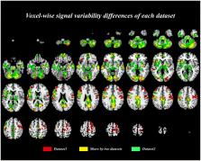
          Cervical spondylotic myelopathy (CSM) is a common disease of the elderly that is characterized by gait instability, sensorimotor deficits, etc. Recurrent symptoms including memory loss, poor attention, etc. have also been reported in recent studies. However, these have been rarely investigated in CSM patients. To investigate the cognitive deficits and their correlation with brain functional alterations, we conducted resting-state fMRI (rs-fMRI) signal variability. This is a novel indicator in the neuroimaging field for assessing the regional neural activity in CSM patients. Further, to explore the network changes in patients, functional connectivity (FC) and graph theory analyses were performed. Compared with the controls, the signal variabilities were significantly lower in the widespread brain regions especially at the default mode network (DMN), visual network, and somatosensory network. The altered inferior parietal lobule signal variability positively correlated with the cognitive function level. Moreover, the FC and the global efficiency of DMN increased in patients with CSM and positively correlated with the cognitive function level. According to the study results, (1) the cervical spondylotic myelopathy patients exhibited regional neural impairments, which correlated with the severity of cognitive deficits in the DMN brain regions, and (2) the increased FC and global efficiency of DMN can compensate for the regional impairment.

          Related collections

          Most cited references85

          • Record: found
          • Abstract: found
          • Article: not found

          Spurious but systematic correlations in functional connectivity MRI networks arise from subject motion.

          Here, we demonstrate that subject motion produces substantial changes in the timecourses of resting state functional connectivity MRI (rs-fcMRI) data despite compensatory spatial registration and regression of motion estimates from the data. These changes cause systematic but spurious correlation structures throughout the brain. Specifically, many long-distance correlations are decreased by subject motion, whereas many short-distance correlations are increased. These changes in rs-fcMRI correlations do not arise from, nor are they adequately countered by, some common functional connectivity processing steps. Two indices of data quality are proposed, and a simple method to reduce motion-related effects in rs-fcMRI analyses is demonstrated that should be flexibly implementable across a variety of software platforms. We demonstrate how application of this technique impacts our own data, modifying previous conclusions about brain development. These results suggest the need for greater care in dealing with subject motion, and the need to critically revisit previous rs-fcMRI work that may not have adequately controlled for effects of transient subject movements. Copyright © 2011 Elsevier Inc. All rights reserved.
            Bookmark
            • Record: found
            • Abstract: not found
            • Article: not found

            Improved Optimization for the Robust and Accurate Linear Registration and Motion Correction of Brain Images

              Bookmark
              • Record: found
              • Abstract: found
              • Article: not found

              The brain's default mode network.

              The brain's default mode network consists of discrete, bilateral and symmetrical cortical areas, in the medial and lateral parietal, medial prefrontal, and medial and lateral temporal cortices of the human, nonhuman primate, cat, and rodent brains. Its discovery was an unexpected consequence of brain-imaging studies first performed with positron emission tomography in which various novel, attention-demanding, and non-self-referential tasks were compared with quiet repose either with eyes closed or with simple visual fixation. The default mode network consistently decreases its activity when compared with activity during these relaxed nontask states. The discovery of the default mode network reignited a longstanding interest in the significance of the brain's ongoing or intrinsic activity. Presently, studies of the brain's intrinsic activity, popularly referred to as resting-state studies, have come to play a major role in studies of the human brain in health and disease. The brain's default mode network plays a central role in this work.
                Bookmark

                Author and article information

                Contributors
                Journal
                Front Neurol
                Front Neurol
                Front. Neurol.
                Frontiers in Neurology
                Frontiers Media S.A.
                1664-2295
                23 December 2020
                2020
                : 11
                : 596795
                Affiliations
                [1] 1Department of Orthopedics Surgery, Tianjin Medical University General Hospital , Tianjin, China
                [2] 2Department of Molecular Imaging and Nuclear Medicine, National Clinical Research Center for Cancer, Tianjin Key Laboratory of Cancer Prevention and Therapy, Tianjin's Clinical Research Center for China, Tianjin Medical University Cancer Institute and Hospital , Tianjin, China
                [3] 3Department of Radiology, Tianjin Medical University General Hospital , Tianjin, China
                [4] 4School of Medical Imaging, Tianjin Medical University , Tianjin, China
                [5] 5Tianjin Key Laboratory of Spine and Spinal Cord, Tianjin Medical University General Hospital , Tianjin, China
                Author notes

                Edited by: Wei Zhang, Peking University First Hospital, China

                Reviewed by: Yashar Zeighami, McGill University, Canada; Daniele Corbo, University of Brescia, Italy

                *Correspondence: Yuan Xue xueyuanzyy@ 123456163.com

                This article was submitted to Applied Neuroimaging, a section of the journal Frontiers in Neurology

                †These authors contributed equally to this work

                Article
                10.3389/fneur.2020.596795
                7785814
                33424749
                b875cb95-52d3-49f8-9c54-ee465c117c57
                Copyright © 2020 Zhao, Su, Chen, Sun, Liang and Xue.

                This is an open-access article distributed under the terms of the Creative Commons Attribution License (CC BY). The use, distribution or reproduction in other forums is permitted, provided the original author(s) and the copyright owner(s) are credited and that the original publication in this journal is cited, in accordance with accepted academic practice. No use, distribution or reproduction is permitted which does not comply with these terms.

                History
                : 20 August 2020
                : 19 November 2020
                Page count
                Figures: 5, Tables: 4, Equations: 0, References: 85, Pages: 15, Words: 9806
                Funding
                Funded by: National Natural Science Foundation of China 10.13039/501100001809
                Award ID: 30973024
                Award ID: 61773223
                Award ID: 81471403
                Award ID: 81871124
                Categories
                Neurology
                Original Research

                Neurology
                resting-state fmri,cervical spondylotic myelopathy,cognitive deficits,bold variability,default mode network

                Comments

                Comment on this article