1
views
0
recommends
+1 Recommend
0 collections
    0
    shares
      • Record: found
      • Abstract: found
      • Article: found
      Is Open Access

      Identification and Therapeutic Outcome Prediction of Cervical Spondylotic Myelopathy Based on the Functional Connectivity From Resting-State Functional MRI Data: A Preliminary Machine Learning Study

      research-article

      Read this article at

      Bookmark
          There is no author summary for this article yet. Authors can add summaries to their articles on ScienceOpen to make them more accessible to a non-specialist audience.

          Abstract

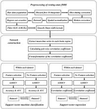
          Currently, strategies to diagnose patients and predict neurological recovery in cervical spondylotic myelopathy (CSM) using MR images of the cervical spine are urgently required. In light of this, this study aimed at exploring potential preoperative brain biomarkers that can be used to diagnose and predict neurological recovery in CSM patients using functional connectivity (FC) analysis of a resting-state functional MRI (rs-fMRI) data. Two independent datasets, including total of 53 patients with CSM and 47 age- and sex-matched healthy controls (HCs), underwent the preoperative rs-fMRI procedure. The FC was calculated from the automated anatomical labeling (AAL) template and used as features for machine learning analysis. After that, three analyses were used, namely, the classification of CSM patients from healthy adults using the support vector machine (SVM) within and across datasets, the prediction of preoperative neurological function in CSM patients via support vector regression (SVR) within and across datasets, and the prediction of neurological recovery in CSM patients via SVR within and across datasets. The results showed that CSM patients could be successfully identified from HCs with high classification accuracies (84.2% for dataset 1, 95.2% for dataset 2, and 73.0% for cross-site validation). Furthermore, the rs-FC combined with SVR could successfully predict the neurological recovery in CSM patients. Additionally, our results from cross-site validation analyses exhibited good reproducibility and generalization across the two datasets. Therefore, our findings provide preliminary evidence toward the development of novel strategies to predict neurological recovery in CSM patients using rs-fMRI and machine learning technique.

          Related collections

          Most cited references50

          • Record: found
          • Abstract: found
          • Article: not found

          Automated anatomical labeling of activations in SPM using a macroscopic anatomical parcellation of the MNI MRI single-subject brain.

          An anatomical parcellation of the spatially normalized single-subject high-resolution T1 volume provided by the Montreal Neurological Institute (MNI) (D. L. Collins et al., 1998, Trans. Med. Imag. 17, 463-468) was performed. The MNI single-subject main sulci were first delineated and further used as landmarks for the 3D definition of 45 anatomical volumes of interest (AVOI) in each hemisphere. This procedure was performed using a dedicated software which allowed a 3D following of the sulci course on the edited brain. Regions of interest were then drawn manually with the same software every 2 mm on the axial slices of the high-resolution MNI single subject. The 90 AVOI were reconstructed and assigned a label. Using this parcellation method, three procedures to perform the automated anatomical labeling of functional studies are proposed: (1) labeling of an extremum defined by a set of coordinates, (2) percentage of voxels belonging to each of the AVOI intersected by a sphere centered by a set of coordinates, and (3) percentage of voxels belonging to each of the AVOI intersected by an activated cluster. An interface with the Statistical Parametric Mapping package (SPM, J. Ashburner and K. J. Friston, 1999, Hum. Brain Mapp. 7, 254-266) is provided as a freeware to researchers of the neuroimaging community. We believe that this tool is an improvement for the macroscopical labeling of activated area compared to labeling assessed using the Talairach atlas brain in which deformations are well known. However, this tool does not alleviate the need for more sophisticated labeling strategies based on anatomical or cytoarchitectonic probabilistic maps.
            Bookmark
            • Record: found
            • Abstract: found
            • Article: not found

            Functional connectivity in the resting brain: a network analysis of the default mode hypothesis.

            Functional imaging studies have shown that certain brain regions, including posterior cingulate cortex (PCC) and ventral anterior cingulate cortex (vACC), consistently show greater activity during resting states than during cognitive tasks. This finding led to the hypothesis that these regions constitute a network supporting a default mode of brain function. In this study, we investigate three questions pertaining to this hypothesis: Does such a resting-state network exist in the human brain? Is it modulated during simple sensory processing? How is it modulated during cognitive processing? To address these questions, we defined PCC and vACC regions that showed decreased activity during a cognitive (working memory) task, then examined their functional connectivity during rest. PCC was strongly coupled with vACC and several other brain regions implicated in the default mode network. Next, we examined the functional connectivity of PCC and vACC during a visual processing task and show that the resultant connectivity maps are virtually identical to those obtained during rest. Last, we defined three lateral prefrontal regions showing increased activity during the cognitive task and examined their resting-state connectivity. We report significant inverse correlations among all three lateral prefrontal regions and PCC, suggesting a mechanism for attenuation of default mode network activity during cognitive processing. This study constitutes, to our knowledge, the first resting-state connectivity analysis of the default mode and provides the most compelling evidence to date for the existence of a cohesive default mode network. Our findings also provide insight into how this network is modulated by task demands and what functions it might subserve.
              Bookmark
              • Record: found
              • Abstract: found
              • Article: not found

              Dynamic functional connectivity: promise, issues, and interpretations.

              The brain must dynamically integrate, coordinate, and respond to internal and external stimuli across multiple time scales. Non-invasive measurements of brain activity with fMRI have greatly advanced our understanding of the large-scale functional organization supporting these fundamental features of brain function. Conclusions from previous resting-state fMRI investigations were based upon static descriptions of functional connectivity (FC), and only recently studies have begun to capitalize on the wealth of information contained within the temporal features of spontaneous BOLD FC. Emerging evidence suggests that dynamic FC metrics may index changes in macroscopic neural activity patterns underlying critical aspects of cognition and behavior, though limitations with regard to analysis and interpretation remain. Here, we review recent findings, methodological considerations, neural and behavioral correlates, and future directions in the emerging field of dynamic FC investigations. Copyright © 2013 Elsevier Inc. All rights reserved.
                Bookmark

                Author and article information

                Contributors
                Journal
                Front Neurol
                Front Neurol
                Front. Neurol.
                Frontiers in Neurology
                Frontiers Media S.A.
                1664-2295
                08 October 2021
                2021
                : 12
                : 711880
                Affiliations
                [1] 1Tianjin Key Laboratory of Cancer Prevention and Therapy, Department of Molecular Imaging and Nuclear Medicine, Tianjin Medical University Cancer Institute and Hospital, National Clinical Research Center for Cancer, Tianjin's Clinical Research Center for China , Tianjin, China
                [2] 2Department of Orthopedics Surgery, Tianjin Medical University General Hospital , Tianjin, China
                [3] 3School of Basic Medical Sciences, Tianjin Medical University , Tianjin, China
                [4] 4School and Hospital of Stomatology, Tianjin Medical University , Tianjin, China
                [5] 5Department of Radiology, Tianjin Medical University General Hospital , Tianjin, China
                Author notes

                Edited by: Marie-Eve Hoeppli, Cincinnati Children's Hospital Medical Center, United States

                Reviewed by: Ailish Malone, Royal College of Surgeons in Ireland, Ireland; Takashi Kaito, Osaka University, Japan

                *Correspondence: Qian Su suqian0510@ 123456126.com

                This article was submitted to Applied Neuroimaging, a section of the journal Frontiers in Neurology

                †These authors have contributed equally to this work

                Article
                10.3389/fneur.2021.711880
                8531403
                34690912
                da4aec01-91d6-4088-bbaf-094b7d37bcb6
                Copyright © 2021 Su, Zhao, Wang, Tu, Guo and Yang.

                This is an open-access article distributed under the terms of the Creative Commons Attribution License (CC BY). The use, distribution or reproduction in other forums is permitted, provided the original author(s) and the copyright owner(s) are credited and that the original publication in this journal is cited, in accordance with accepted academic practice. No use, distribution or reproduction is permitted which does not comply with these terms.

                History
                : 19 May 2021
                : 19 August 2021
                Page count
                Figures: 6, Tables: 2, Equations: 2, References: 50, Pages: 14, Words: 9163
                Categories
                Neurology
                Original Research

                Neurology
                rs-fmri,machine learning,cervical spondylotic myelopathy,support vector machine,functional connectivity

                Comments

                Comment on this article