2
views
0
recommends
+1 Recommend
0 collections
    0
    shares
      • Record: found
      • Abstract: found
      • Article: found
      Is Open Access

      Remodeling of the brain correlates with gait instability in cervical spondylotic myelopathy

      research-article

      Read this article at

      Bookmark
          There is no author summary for this article yet. Authors can add summaries to their articles on ScienceOpen to make them more accessible to a non-specialist audience.

          Abstract

          Introduction

          Cervical spondylotic myelopathy (CSM) is a common form of non-traumatic spinal cord injury (SCI) and usually leads to remodeling of the brain and spinal cord. In CSM with gait instability, the remodeling of the brain and cervical spinal cord is unclear. We attempted to explore the remodeling of these patients’ brains and spinal cords, as well as the relationship between the remodeling of the brain and spinal cord and gait instability.

          Methods

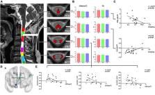
          According to the CSM patients’ gait, we divided patients into two groups: normal gait patients (nPT) and abnormal gait patients (aPT). Voxel-wise z-score transformation amplitude of low-frequency fluctuations (zALFF) and resting-state functional connectivity (rs-FC) were performed for estimating brain changes. Cross-sectional area (CSA) and fractional anisotropy (FA) of the spinal cord were computed by Spinal cord toolbox. Correlations of these measures and the modified Japanese Orthopedic Association (mJOA) score were analyzed.

          Results

          We found that the zALFF of caudate nucleus in aPT was higher than that in healthy controls (HC) and lower than that in nPT. The zALFF of the right postcentral gyrus and paracentral lobule in HC was higher than those of aPT and nPT. Compared with the nPT, the aPT showed increased functional connectivity between the caudate nucleus and left angular gyrus, bilateral precuneus and bilateral posterior cingulate cortex (PCC), which constitute a vital section of the default mode network (DMN). No significantly different FA values or CSA of spinal tracts at the C2 level were observed between the HC, nPT and aPT groups. In CSM, the right paracentral lobule’s zALFF was negatively correlated with the FA value of fasciculus gracilis (FCG), and the right caudate zALFF was positively correlated with the FA value of the fasciculus cuneatus (FCC). The results showed that the functional connectivity between the right caudate nucleus and DMN was negatively correlated with the CSA of the lateral corticospinal tract (CST).

          Discussion

          The activation of the caudate nucleus and the strengthening functional connectivity between the caudate nucleus and DMN were associated with gait instability in CSM patients. Correlations between spinal cord and brain function might be related to the clinical symptoms in CSM.

          Related collections

          Most cited references47

          • Record: found
          • Abstract: found
          • Article: not found

          Decoding the organization of spinal circuits that control locomotion.

          Ole Kiehn (2016)
          Unravelling the functional operation of neuronal networks and linking cellular activity to specific behavioural outcomes are among the biggest challenges in neuroscience. In this broad field of research, substantial progress has been made in studies of the spinal networks that control locomotion. Through united efforts using electrophysiological and molecular genetic network approaches and behavioural studies in phylogenetically diverse experimental models, the organization of locomotor networks has begun to be decoded. The emergent themes from this research are that the locomotor networks have a modular organization with distinct transmitter and molecular codes and that their organization is reconfigured with changes to the speed of locomotion or changes in gait.
            Bookmark
            • Record: found
            • Abstract: not found
            • Article: not found

            The default mode network in cognition: a topographical perspective

              Bookmark
              • Record: found
              • Abstract: found
              • Article: not found

              SCT: Spinal Cord Toolbox, an open-source software for processing spinal cord MRI data

              For the past 25 years, the field of neuroimaging has witnessed the development of several software packages for processing multi-parametric magnetic resonance imaging (mpMRI) to study the brain. These software packages are now routinely used by researchers and clinicians, and have contributed to important breakthroughs for the understanding of brain anatomy and function. However, no software package exists to process mpMRI data of the spinal cord. Despite the numerous clinical needs for such advanced mpMRI protocols (multiple sclerosis, spinal cord injury, cervical spondylotic myelopathy, etc.), researchers have been developing specific tools that, while necessary, do not provide an integrative framework that is compatible with most usages and that is capable of reaching the community at large. This hinders cross-validation and the possibility to perform multi-center studies. In this study we introduce the Spinal Cord Toolbox (SCT), a comprehensive software dedicated to the processing of spinal cord MRI data. SCT builds on previously-validated methods and includes state-of-the-art MRI templates and atlases of the spinal cord, algorithms to segment and register new data to the templates, and motion correction methods for diffusion and functional time series. SCT is tailored towards standardization and automation of the processing pipeline, versatility, modularity, and it follows guidelines of software development and distribution. Preliminary applications of SCT cover a variety of studies, from cross-sectional area measures in large databases of patients, to the precise quantification of mpMRI metrics in specific spinal pathways. We anticipate that SCT will bring together the spinal cord neuroimaging community by establishing standard templates and analysis procedures.
                Bookmark

                Author and article information

                Contributors
                Journal
                Front Neurosci
                Front Neurosci
                Front. Neurosci.
                Frontiers in Neuroscience
                Frontiers Media S.A.
                1662-4548
                1662-453X
                02 February 2023
                2023
                : 17
                : 1087945
                Affiliations
                [1] 1Department of Orthopedics, The First Affiliated Hospital of Anhui Medical University , Hefei, China
                [2] 2Department of Spine Surgery, The First Affiliated Hospital of Anhui Medical University , Hefei, China
                [3] 3Department of Radiology, The First Affiliated Hospital of Anhui Medical University , Hefei, China
                [4] 4School of Basic Medical Sciences, Anhui Medical University , Hefei, China
                [5] 5Department of Orthopedics, Dongcheng Branch of The First Affiliated Hospital of Anhui Medical University (Feidong People’s Hospital) , Hefei, China
                [6] 6Department of Medical Imaging, The First Affiliated Hospital of Anhui Medical University , Hefei, China
                Author notes

                Edited by: Andrew S. Nencka, Medical College of Wisconsin, United States

                Reviewed by: Peter Pahapill, Medical College of Wisconsin, United States; Jia-Sheng Rao, Beihang University, China

                *Correspondence: Fulong Dong, dongfulongtg@ 123456sina.com

                These authors have contributed equally to this work and share first authorship

                These authors have contributed equally to this work

                This article was submitted to Brain Imaging Methods, a section of the journal Frontiers in Neuroscience

                Article
                10.3389/fnins.2023.1087945
                9932596
                36816111
                2561f11f-e2c9-4123-b491-d69239c312d2
                Copyright © 2023 Wu, Wang, Chang, Zhu, Zhang, Li, Zuo, Chen, Jin, Yan, Yang, Xu, Song, Wu, Qian, Shen, Yu and Dong.

                This is an open-access article distributed under the terms of the Creative Commons Attribution License (CC BY). The use, distribution or reproduction in other forums is permitted, provided the original author(s) and the copyright owner(s) are credited and that the original publication in this journal is cited, in accordance with accepted academic practice. No use, distribution or reproduction is permitted which does not comply with these terms.

                History
                : 17 November 2022
                : 17 January 2023
                Page count
                Figures: 5, Tables: 3, Equations: 0, References: 47, Pages: 10, Words: 7194
                Funding
                Funded by: University Natural Science Research Project of Anhui Province , doi 10.13039/501100009558;
                Award ID: Grant No. KJ2019A0275
                Funded by: Anhui Provincial Quality Engineering Project , doi 10.13039/100015801;
                Award ID: Grant No. 2020jyxm0918
                This work was supported by University Research Foundation of Anhui Medical University (Grant No. 2020xkj175), The First Affiliated Hospital of Anhui Medical University Clinical Research Project (Grant No. LCYJ2021YB018), and University Natural Science Research Key Project of Anhui Province (Grant No. 2022AH051158).
                Categories
                Neuroscience
                Original Research

                Neurosciences
                resting-state fmri,diffusion tensor imaging,cervical spondylotic myelopathy,gait instability,caudate nucleus,corticospinal tract

                Comments

                Comment on this article