41
views
0
recommends
+1 Recommend
0 collections
    0
    shares
      • Record: found
      • Abstract: found
      • Article: found
      Is Open Access

      Predictive value of in vitro assays depends on the mechanism of toxicity of metal oxide nanoparticles

      research-article

      Read this article at

      Bookmark
          There is no author summary for this article yet. Authors can add summaries to their articles on ScienceOpen to make them more accessible to a non-specialist audience.

          Abstract

          Background

          Hazard identification for risk assessment of nanoparticles (NPs) is mainly composed of in vitro cell-based assays and in vivo animal experimentation. The rapidly increasing number and functionalizations of NPs makes in vivo toxicity tests undesirable on both ethical and financial grounds, creating an urgent need for development of in vitro cell-based assays that accurately predict in vivo toxicity and facilitate safe nanotechnology.

          Methods

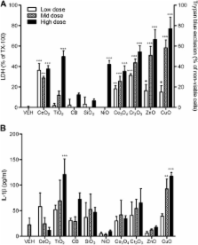
          In this study, we used 9 different NPs (CeO 2, TiO 2, carbon black, SiO 2, NiO, Co 3O 4, Cr 2O 3, CuO, and ZnO). As an in vivo toxicity endpoint, the acute lung inflammogenicity in a rat instillation model was compared with the in vitro toxicity endpoints comprising cytotoxicity, pro-inflammatory cytokine expression, or haemolytic potential. For in vitro assays, 8 different cell-based assays were used including epithelial cells, monocytic/macrophage cells, human erythrocytes, and combined culture.

          Results

          ZnO and CuO NPs acting via soluble toxic ions showed positive results in most of assays and were consistent with the lung inflammation data. When compared in in vitro assays at the same surface area dose (30 cm 2/mL), NPs that were low solubility and therefore acting via surface reactivity had no convincing activity, except for CeO 2 NP. Cytotoxicity in differentiated peripheral blood mononuclear cells was the most accurate showing 89% accuracy and 11% false negativity in predicting acute lung inflammogenicity. However, the haemolysis assay showed 100% consistency with the lung inflammation if any dose, having statistical significance was considered positivity. Other cell-based in vitro assays showed a poorer correlation with in vivo inflammogenicity.

          Conclusions

          Based on the toxicity mechanisms of NPs, two different approaches can be applied for prediction of in vivo lung inflammogenicity. Most in vitro assays were good at detecting NPs that act via soluble ions (i.e., ZnO and CuO NP). However, in vitro assays were limited in detecting NPs acting via surface reactivity as their mechanism of toxicity, except for the haemolysis assay.

          Related collections

          Most cited references30

          • Record: found
          • Abstract: found
          • Article: found
          Is Open Access

          Principles for characterizing the potential human health effects from exposure to nanomaterials: elements of a screening strategy

          The rapid proliferation of many different engineered nanomaterials (defined as materials designed and produced to have structural features with at least one dimension of 100 nanometers or less) presents a dilemma to regulators regarding hazard identification. The International Life Sciences Institute Research Foundation/Risk Science Institute convened an expert working group to develop a screening strategy for the hazard identification of engineered nanomaterials. The working group report presents the elements of a screening strategy rather than a detailed testing protocol. Based on an evaluation of the limited data currently available, the report presents a broad data gathering strategy applicable to this early stage in the development of a risk assessment process for nanomaterials. Oral, dermal, inhalation, and injection routes of exposure are included recognizing that, depending on use patterns, exposure to nanomaterials may occur by any of these routes. The three key elements of the toxicity screening strategy are: Physicochemical Characteristics, In Vitro Assays (cellular and non-cellular), and In Vivo Assays. There is a strong likelihood that biological activity of nanoparticles will depend on physicochemical parameters not routinely considered in toxicity screening studies. Physicochemical properties that may be important in understanding the toxic effects of test materials include particle size and size distribution, agglomeration state, shape, crystal structure, chemical composition, surface area, surface chemistry, surface charge, and porosity. In vitro techniques allow specific biological and mechanistic pathways to be isolated and tested under controlled conditions, in ways that are not feasible in in vivo tests. Tests are suggested for portal-of-entry toxicity for lungs, skin, and the mucosal membranes, and target organ toxicity for endothelium, blood, spleen, liver, nervous system, heart, and kidney. Non-cellular assessment of nanoparticle durability, protein interactions, complement activation, and pro-oxidant activity is also considered. Tier 1 in vivo assays are proposed for pulmonary, oral, skin and injection exposures, and Tier 2 evaluations for pulmonary exposures are also proposed. Tier 1 evaluations include markers of inflammation, oxidant stress, and cell proliferation in portal-of-entry and selected remote organs and tissues. Tier 2 evaluations for pulmonary exposures could include deposition, translocation, and toxicokinetics and biopersistence studies; effects of multiple exposures; potential effects on the reproductive system, placenta, and fetus; alternative animal models; and mechanistic studies.
            Bookmark
            • Record: found
            • Abstract: not found
            • Article: not found

            A review of the in vivo and in vitro toxicity of silver and gold particulates: particle attributes and biological mechanisms responsible for the observed toxicity.

            This review is concerned with evaluating the toxicity associated with human exposure to silver and gold nanoparticles (NPs), due to the relative abundance of toxicity data available for these particles, when compared to other metal particulates. This has allowed knowledge on the current understanding of the field to be gained, and has demonstrated where gaps in knowledge are. It is anticipated that evaluating the hazards associated with silver and gold particles will ultimately enable risk assessments to be completed, by combining this information with knowledge on the level of human exposure. The quantity of available hazard information for metals is greatest for silver particulates, due to its widespread inclusion within a number of diverse products (including clothes and wound dressings), which primarily arises from its antibacterial behaviour. Gold has been used on numerous occasions to assess the biodistribution and cellular uptake of NPs following exposure. Inflammatory, oxidative, genotoxic, and cytotoxic consequences are associated with silver particulate exposure, and are inherently linked. The primary site of gold and silver particulate accumulation has been consistently demonstrated to be the liver, and it is therefore relevant that a number of in vitro investigations have focused on this potential target organ. However, in general there is a lack of in vivo and in vitro toxicity information that allows correlations between the findings to be made. Instead a focus on the tissue distribution of particles following exposure is evident within the available literature, which can be useful in directing appropriate in vitro experimentation by revealing potential target sites of toxicity. The experimental design has the potential to impact on the toxicological observations, and in particular the use of excessively high particle concentrations has been observed. As witnessed for other particle types, gold and silver particle sizes are influential in dictating the observed toxicity, with smaller particles exhibiting a greater response than their larger counterparts, and this is likely to be driven by differences in particle surface area, when administered at an equal-mass dose. A major obstacle, at present, is deciphering whether the responses related to silver nanoparticulate exposure derive from their small size, or particle dissolution contributes to the observed toxicity. Alternatively, a combination of both may be responsible, as the release of ions would be expected to be greater for smaller particles.
              Bookmark
              • Record: found
              • Abstract: found
              • Article: not found

              Optimized THP-1 differentiation is required for the detection of responses to weak stimuli.

              The differentiation of THP-1 monocytes into macrophages is mainly conducted at a phorbol 12-myristate 13-acetate (PMA) concentration of 10-400 ng/ml. However, this concentration might be high enough to upregulate the expressions of some genes in differentiated macrophages, which could overwhelm gene expression increases induced by other stimuli. The present study was performed to optimize the PMA concentration required to differentiate monocytes whilst minimizing gene upregulation. THP-1 cells were treated with 2.5-100 ng/ml PMA and analyzed for the extent of cell adherence, the surface marker of macrophages, and stable differentiation without undesirable gene upregulation. The stably differentiated THP-1 cells at the minimum PMA concentration were treated with 10 ng/ml LPS or 125 nM amyloid beta (Abeta(1-42)). The treatment of THP-1 with 5 ng/ml PMA was found to be sufficient to induce stable differentiation without undesirable gene upregulation. These macrophages differentiated at 5 ng/ml responded well to secondary weak stimuli like 10 ng/ml LPS or 125 nM of amyloid beta (Abeta(1-42)). This finding suggests that THP-1 cells are well differentiated by 5 ng/ml PMA, and that the resulting differentiated macrophages respond well to secondary weak stimuli without being overwhelmed by undesirable gene upregulation induced by PMA.
                Bookmark

                Author and article information

                Contributors
                Journal
                Part Fibre Toxicol
                Part Fibre Toxicol
                Particle and Fibre Toxicology
                BioMed Central
                1743-8977
                2013
                25 October 2013
                : 10
                : 55
                Affiliations
                [1 ]ELEGI/Colt Laboratory, Centre for Inflammation Research, University of Edinburgh, 47 Little France Crescent, Edinburgh EH16 4TJ, UK
                [2 ]Department of Medicinal Biotechnology, College of Natural Resources and Life Science, Dong-A University, Busan 604-714, Republic of Korea
                [3 ]School of Chemistry, West Mains Road, University of Edinburgh, Edinburgh, UK
                [4 ]Free Radical Research Facility, Department of Diabetes & Cardiovascular Science, University of the Highlands & Islands, Centre for Health Science, Inverness, UK
                [5 ]Department of Toxicological Research, National Institute of Food and Drug Safety Evaluation, Ministry of Food and Drug Safety, Osong 363-700, Republic of Korea
                Article
                1743-8977-10-55
                10.1186/1743-8977-10-55
                4016420
                24156363
                af4d5e3c-8da3-4c9f-923c-bbbd0353e8ea
                Copyright © 2013 Cho et al.; licensee BioMed Central Ltd.

                This is an open access article distributed under the terms of the Creative Commons Attribution License ( http://creativecommons.org/licenses/by/2.0), which permits unrestricted use, distribution, and reproduction in any medium, provided the original work is properly cited.

                History
                : 8 April 2013
                : 21 October 2013
                Categories
                Research

                Toxicology
                in vitro,in vivo,inflammation,mechanism,nanoparticles,prediction,toxicity
                Toxicology
                in vitro, in vivo, inflammation, mechanism, nanoparticles, prediction, toxicity

                Comments

                Comment on this article