26
views
0
recommends
+1 Recommend
0 collections
    0
    shares
      • Record: found
      • Abstract: found
      • Article: found
      Is Open Access

      Analysis of enamel development using murine model systems: approaches and limitations

      review-article

      Read this article at

      Bookmark
          There is no author summary for this article yet. Authors can add summaries to their articles on ScienceOpen to make them more accessible to a non-specialist audience.

          Abstract

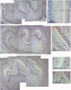
          A primary goal of enamel research is to understand and potentially treat or prevent enamel defects related to amelogenesis imperfecta (AI). Rodents are ideal models to assist our understanding of how enamel is formed because they are easily genetically modified, and their continuously erupting incisors display all stages of enamel development and mineralization. While numerous methods have been developed to generate and analyze genetically modified rodent enamel, it is crucial to understand the limitations and challenges associated with these methods in order to draw appropriate conclusions that can be applied translationally, to AI patient care. We have highlighted methods involved in generating and analyzing rodent enamel and potential approaches to overcoming limitations of these methods: (1) generating transgenic, knockout, and knockin mouse models, and (2) analyzing rodent enamel mineral density and functional properties (structure and mechanics) of mature enamel. There is a need for a standardized workflow to analyze enamel phenotypes in rodent models so that investigators can compare data from different studies. These methods include analyses of gene and protein expression, developing enamel histology, enamel pigment, degree of mineralization, enamel structure, and mechanical properties. Standardization of these methods with regard to stage of enamel development and sample preparation is crucial, and ideally investigators can use correlative and complementary techniques with the understanding that developing mouse enamel is dynamic and complex.

          Related collections

          Most cited references59

          • Record: found
          • Abstract: found
          • Article: not found

          Disruption of the proto-oncogene int-2 in mouse embryo-derived stem cells: a general strategy for targeting mutations to non-selectable genes.

          Gene targeting--homologous recombination of DNA sequences residing in the chromosome with newly introduced DNA sequences--in mouse embryo-derived stem cells promises to provide a means to generate mice of any desired genotype. We describe a positive nd negative selection procedure that enriches 2,000-fold for those cells that contain a targeted mutation. The procedure was applied to the isolation of hprt- and int-2- mutants, but it should be applicable to any gene.
            Bookmark
            • Record: found
            • Abstract: found
            • Article: found
            Is Open Access

            Iron Deposition and Ferritin Heavy Chain (Fth) Localization in Rodent Teeth

            Background An iron rich layer on the labial surface is characteristic of the enamel of rodent incisors. In order to address a role for iron content in continuously growing incisors during odontogenesis, we studied iron deposition patterns in enamel and dentine using Perls’ blue staining and ferritin heavy chain (Fth) immunolocalization. Fth expression is regulated by iron level; therefore its localization can be used as a sensitive indicator for iron deposition. Results Sagittal sections of 4-week old rat incisors showed a gradual increase in iron level in the enamel organ from secretory to maturation stages. In addition, iron was detected in ameloblasts of erupting third molars of 4-week old rats, suggesting iron plays a role in both incisor and molar development. In odontoblasts, the presence of iron was demonstrated, and this is consistent with iron’s role in collagen synthesis. Using postnatal 3-, 6-, 9-day old mice, the spatial and temporal expression of Fth in tooth development again indicated the presence of iron in mature ameloblasts and odontoblasts. Conclusions While these data do not explain what functional role iron has in tooth formation, it does highlight a significant molecular activity associated with the formation of the rodent dentition.
              Bookmark
              • Record: found
              • Abstract: found
              • Article: found
              Is Open Access

              Dental Enamel Development: Proteinases and Their Enamel Matrix Substrates

              This review focuses on recent discoveries and delves in detail about what is known about each of the proteins (amelogenin, ameloblastin, and enamelin) and proteinases (matrix metalloproteinase-20 and kallikrein-related peptidase-4) that are secreted into the enamel matrix. After an overview of enamel development, this review focuses on these enamel proteins by describing their nomenclature, tissue expression, functions, proteinase activation, and proteinase substrate specificity. These proteins and their respective null mice and human mutations are also evaluated to shed light on the mechanisms that cause nonsyndromic enamel malformations termed amelogenesis imperfecta. Pertinent controversies are addressed. For example, do any of these proteins have a critical function in addition to their role in enamel development? Does amelogenin initiate crystallite growth, does it inhibit crystallite growth in width and thickness, or does it do neither? Detailed examination of the null mouse literature provides unmistakable clues and/or answers to these questions, and this data is thoroughly analyzed. Striking conclusions from this analysis reveal that widely held paradigms of enamel formation are inadequate. The final section of this review weaves the recent data into a plausible new mechanism by which these enamel matrix proteins support and promote enamel development.
                Bookmark

                Author and article information

                Contributors
                Journal
                Front Physiol
                Front Physiol
                Front. Physiol.
                Frontiers in Physiology
                Frontiers Media S.A.
                1664-042X
                17 September 2014
                2014
                : 5
                : 313
                Affiliations
                [1] 1Department of Mineralized Tissue Biology, The Forsyth Institute, Harvard School of Dental Medicine, Harvard University Cambridge, MA, USA
                [2] 2Department of Anatomy and Cell Biology, School of Dental Medicine, University of Pennsylvania Philadelphia, PA, USA
                Author notes

                Edited by: Bernhard Ganss, University of Toronto, Canada

                Reviewed by: Harald Osmundsen, University of Oslo, Norway; Catherine Chaussain, Université Paris Descartes Paris Cité, France

                *Correspondence: Megan K. Pugach, Department of Mineralized Tissue Biology, Harvard School of Dental Medicine, The Forsyth Institute, 245 First Street, Cambridge, MA 02142, USA e-mail: mpugach@ 123456forsyth.org

                This article was submitted to Craniofacial Biology, a section of the journal Frontiers in Physiology.

                Article
                10.3389/fphys.2014.00313
                4166228
                25278900
                a65f46e2-38d9-470a-8ccf-3a075be22c4e
                Copyright © 2014 Pugach and Gibson.

                This is an open-access article distributed under the terms of the Creative Commons Attribution License (CC BY). The use, distribution or reproduction in other forums is permitted, provided the original author(s) or licensor are credited and that the original publication in this journal is cited, in accordance with accepted academic practice. No use, distribution or reproduction is permitted which does not comply with these terms.

                History
                : 16 June 2014
                : 01 August 2014
                Page count
                Figures: 4, Tables: 1, Equations: 0, References: 70, Pages: 10, Words: 8175
                Categories
                Physiology
                Review Article

                Anatomy & Physiology
                enamel development,transgenic,knockout,knockin,amelogenin,mineralization
                Anatomy & Physiology
                enamel development, transgenic, knockout, knockin, amelogenin, mineralization

                Comments

                Comment on this article