6
views
0
recommends
+1 Recommend
1 collections
    0
    shares
      • Record: found
      • Abstract: found
      • Article: found
      Is Open Access

      Harnessing Machine Learning in Early COVID-19 Detection and Prognosis: A Comprehensive Systematic Review

      review-article

      Read this article at

      Bookmark
          There is no author summary for this article yet. Authors can add summaries to their articles on ScienceOpen to make them more accessible to a non-specialist audience.

          Abstract

          During the early phase of the COVID-19 pandemic, reverse transcriptase-polymerase chain reaction (RT-PCR) testing faced limitations, prompting the exploration of machine learning (ML) alternatives for diagnosis and prognosis. Providing a comprehensive appraisal of such decision support systems and their use in COVID-19 management can aid the medical community in making informed decisions during the risk assessment of their patients, especially in low-resource settings. Therefore, the objective of this study was to systematically review the studies that predicted the diagnosis of COVID-19 or the severity of the disease using ML.

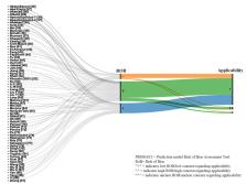
          Following the Preferred Reporting Items for Systematic Reviews and Meta-Analysis (PRISMA), we conducted a literature search of MEDLINE (OVID), Scopus, EMBASE, and IEEE Xplore from January 1 to June 31, 2020. The outcomes were COVID-19 diagnosis or prognostic measures such as death, need for mechanical ventilation, admission, and acute respiratory distress syndrome. We included peer-reviewed observational studies, clinical trials, research letters, case series, and reports. We extracted data about the study's country, setting, sample size, data source, dataset, diagnostic or prognostic outcomes, prediction measures, type of ML model, and measures of diagnostic accuracy. Bias was assessed using the Prediction model Risk Of Bias ASsessment Tool (PROBAST). This study was registered in the International Prospective Register of Systematic Reviews (PROSPERO), with the number CRD42020197109.

          The final records included for data extraction were 66. Forty-three (64%) studies used secondary data. The majority of studies were from Chinese authors (30%). Most of the literature (79%) relied on chest imaging for prediction, while the remainder used various laboratory indicators, including hematological, biochemical, and immunological markers. Thirteen studies explored predicting COVID-19 severity, while the rest predicted diagnosis. Seventy percent of the articles used deep learning models, while 30% used traditional ML algorithms. Most studies reported high sensitivity, specificity, and accuracy for the ML models (exceeding 90%). The overall concern about the risk of bias was "unclear" in 56% of the studies. This was mainly due to concerns about selection bias.

          ML may help identify COVID-19 patients in the early phase of the pandemic, particularly in the context of chest imaging. Although these studies reflect that these ML models exhibit high accuracy, the novelty of these models and the biases in dataset selection make using them as a replacement for the clinicians' cognitive decision-making questionable. Continued research is needed to enhance the robustness and reliability of ML systems in COVID-19 diagnosis and prognosis.

          Related collections

          Most cited references106

          • Record: found
          • Abstract: found
          • Article: not found

          Correlation of Chest CT and RT-PCR Testing in Coronavirus Disease 2019 (COVID-19) in China: A Report of 1014 Cases

          Background Chest CT is used for diagnosis of 2019 novel coronavirus disease (COVID-19), as an important complement to the reverse-transcription polymerase chain reaction (RT-PCR) tests. Purpose To investigate the diagnostic value and consistency of chest CT as compared with comparison to RT-PCR assay in COVID-19. Methods From January 6 to February 6, 2020, 1014 patients in Wuhan, China who underwent both chest CT and RT-PCR tests were included. With RT-PCR as reference standard, the performance of chest CT in diagnosing COVID-19 was assessed. Besides, for patients with multiple RT-PCR assays, the dynamic conversion of RT-PCR results (negative to positive, positive to negative, respectively) was analyzed as compared with serial chest CT scans for those with time-interval of 4 days or more. Results Of 1014 patients, 59% (601/1014) had positive RT-PCR results, and 88% (888/1014) had positive chest CT scans. The sensitivity of chest CT in suggesting COVID-19 was 97% (95%CI, 95-98%, 580/601 patients) based on positive RT-PCR results. In patients with negative RT-PCR results, 75% (308/413) had positive chest CT findings; of 308, 48% were considered as highly likely cases, with 33% as probable cases. By analysis of serial RT-PCR assays and CT scans, the mean interval time between the initial negative to positive RT-PCR results was 5.1 ± 1.5 days; the initial positive to subsequent negative RT-PCR result was 6.9 ± 2.3 days). 60% to 93% of cases had initial positive CT consistent with COVID-19 prior (or parallel) to the initial positive RT-PCR results. 42% (24/57) cases showed improvement in follow-up chest CT scans before the RT-PCR results turning negative. Conclusion Chest CT has a high sensitivity for diagnosis of COVID-19. Chest CT may be considered as a primary tool for the current COVID-19 detection in epidemic areas. A translation of this abstract in Farsi is available in the supplement. - ترجمه چکیده این مقاله به فارسی، در ضمیمه موجود است.
            Bookmark
            • Record: found
            • Abstract: found
            • Article: not found

            Automated detection of COVID-19 cases using deep neural networks with X-ray images

            The novel coronavirus 2019 (COVID-2019), which first appeared in Wuhan city of China in December 2019, spread rapidly around the world and became a pandemic. It has caused a devastating effect on both daily lives, public health, and the global economy. It is critical to detect the positive cases as early as possible so as to prevent the further spread of this epidemic and to quickly treat affected patients. The need for auxiliary diagnostic tools has increased as there are no accurate automated toolkits available. Recent findings obtained using radiology imaging techniques suggest that such images contain salient information about the COVID-19 virus. Application of advanced artificial intelligence (AI) techniques coupled with radiological imaging can be helpful for the accurate detection of this disease, and can also be assistive to overcome the problem of a lack of specialized physicians in remote villages. In this study, a new model for automatic COVID-19 detection using raw chest X-ray images is presented. The proposed model is developed to provide accurate diagnostics for binary classification (COVID vs. No-Findings) and multi-class classification (COVID vs. No-Findings vs. Pneumonia). Our model produced a classification accuracy of 98.08% for binary classes and 87.02% for multi-class cases. The DarkNet model was used in our study as a classifier for the you only look once (YOLO) real time object detection system. We implemented 17 convolutional layers and introduced different filtering on each layer. Our model (available at (https://github.com/muhammedtalo/COVID-19)) can be employed to assist radiologists in validating their initial screening, and can also be employed via cloud to immediately screen patients.
              Bookmark
              • Record: found
              • Abstract: found
              • Article: not found

              Covid-19: automatic detection from X-ray images utilizing transfer learning with convolutional neural networks

              In this study, a dataset of X-ray images from patients with common bacterial pneumonia, confirmed Covid-19 disease, and normal incidents, was utilized for the automatic detection of the Coronavirus disease. The aim of the study is to evaluate the performance of state-of-the-art convolutional neural network architectures proposed over the recent years for medical image classification. Specifically, the procedure called Transfer Learning was adopted. With transfer learning, the detection of various abnormalities in small medical image datasets is an achievable target, often yielding remarkable results. The datasets utilized in this experiment are two. Firstly, a collection of 1427 X-ray images including 224 images with confirmed Covid-19 disease, 700 images with confirmed common bacterial pneumonia, and 504 images of normal conditions. Secondly, a dataset including 224 images with confirmed Covid-19 disease, 714 images with confirmed bacterial and viral pneumonia, and 504 images of normal conditions. The data was collected from the available X-ray images on public medical repositories. The results suggest that Deep Learning with X-ray imaging may extract significant biomarkers related to the Covid-19 disease, while the best accuracy, sensitivity, and specificity obtained is 96.78%, 98.66%, and 96.46% respectively. Since by now, all diagnostic tests show failure rates such as to raise concerns, the probability of incorporating X-rays into the diagnosis of the disease could be assessed by the medical community, based on the findings, while more research to evaluate the X-ray approach from different aspects may be conducted.
                Bookmark

                Author and article information

                Journal
                Cureus
                Cureus
                2168-8184
                Cureus
                Cureus (Palo Alto (CA) )
                2168-8184
                1 May 2023
                May 2023
                : 15
                : 5
                : e38373
                Affiliations
                [1 ] Family & Community Medicine Department, College of Medicine, King Saud University, Riyadh, SAU
                [2 ] Research Chair for Evidence-Based Health Care and Knowledge Translation, Family and Community Medicine Department, College of Medicine, King Saud University, Riyadh, SAU
                [3 ] Health Informatics, Public Health Informatics Foundation, Dhaka, BGD
                [4 ] Quality Management Department, King Saud University Medical City, Riyadh, SAU
                [5 ] Pediatrics, Quality Management Department, King Saud University Medical City, Riyadh, SAU
                [6 ] Health Information Management Department, Prince Sultan Military College of Health Sciences, Al Dhahran, SAU
                [7 ] Medicine, Wegner Health Sciences Library, University of South Dakota, Vermillion, USA
                [8 ] Department of Information Systems, King Abdulaziz University, Jeddah, SAU
                [9 ] Department of Radiology, King Saud University, Riyadh, SAU
                [10 ] School of Computer Sciences, Universiti Sains Malaysia, Penang, MYS
                [11 ] Pediatric Intensive Care Unit, Department of Pediatrics, King Saud University, Riyadh, SAU
                Author notes
                Article
                10.7759/cureus.38373
                10230599
                9cbb560e-a71a-4fe1-9d06-70f63ceacbd9
                Copyright © 2023, Dabbagh et al.

                This is an open access article distributed under the terms of the Creative Commons Attribution License, which permits unrestricted use, distribution, and reproduction in any medium, provided the original author and source are credited.

                History
                : 30 April 2023
                Categories
                Radiology
                Infectious Disease
                Healthcare Technology

                healthcare technology,deep learning artificial intelligence,covid-19 chest imaging,decision support systems,covid-19 diagnosis,prediction,artificial intelligence,machine learning in early pandemic,sars-cov-2,covid-19

                Comments

                Comment on this article