7
views
0
recommends
+1 Recommend
0 collections
    0
    shares
      • Record: found
      • Abstract: found
      • Article: found
      Is Open Access

      Fucosylated oligosaccharides in mother’s milk alleviate the effects of caesarean birth on infant gut microbiota

      research-article

      Read this article at

      Bookmark
          There is no author summary for this article yet. Authors can add summaries to their articles on ScienceOpen to make them more accessible to a non-specialist audience.

          Abstract

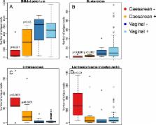
          One of the most abundant components in human milk is formed by oligosaccharides, which are poorly digested by the infant. The oligosaccharide composition of breast milk varies between mothers, and is dependent on maternal secretor (FUT2) genotype. Secretor mothers produce milk containing α1-2 fucosylated human milk oligosaccharides, which are absent in the milk of non-secretor mothers. Several strains of bacteria in the infant gut have the capacity to utilise human milk oligosaccharides (HMOs). Here we investigate the differences in infant gut microbiota composition between secretor (N = 76) and non-secretor (N = 15) mothers, taking into account birth mode. In the vaginally born infants, maternal secretor status was not associated with microbiota composition. In the caesarean-born, however, many of the caesarean-associated microbiota patterns were more pronounced among the infants of non-secretor mothers compared to those of secretor mothers. Particularly bifidobacteria were strongly depleted and enterococci increased among the caesarean-born infants of non-secretor mothers. Furthermore, Akkermansia was increased in the section-born infants of secretor mothers, supporting the suggestion that this organism may degrade HMOs. The results indicate that maternal secretor status may be particularly influential in infants with compromised microbiota development, and that these infants could benefit from corrective supplementation.

          Related collections

          Most cited references32

          • Record: found
          • Abstract: found
          • Article: not found

          Campylobacter jejuni binds intestinal H(O) antigen (Fuc alpha 1, 2Gal beta 1, 4GlcNAc), and fucosyloligosaccharides of human milk inhibit its binding and infection.

          The most common cause of infant mortality is diarrhea; the most common cause of bacterial diarrhea is Campylobacter jejuni, which is also the primary cause of motor neuron paralysis. The first step in campylobacter pathogenesis is adherence to intestinal mucosa. We found that such binding was inhibited in vitro by human milk and, with high avidity, by alpha1,2-fucosylated carbohydrate moieties containing the H(O) blood group epitope (Fuc alpha 1,2Gal beta 1,4GlcNAc em leader ). In studies on the mechanism of adherence, campylobacter, which normally does not bind to Chinese hamster ovary cells, bound avidly when the cells were transfected with a human alpha1,2-fucosyltransferase gene that caused overexpression of H-2 antigen; binding was specifically inhibited by H-2 ligands (lectins Ulex europaeus and Lotus tetragonolobus and H-2 monoclonal antibody), H-2 mimetics, and human milk oligosaccharides. Human milk oligosaccharides inhibited campylobacter colonization of mice in vivo and human intestinal mucosa ex vivo. Campylobacter colonization of nursing mouse pups was inhibited if their dams had been transfected with a human alpha1,2-fucosyltransferase gene that caused expression of H(O) antigen in milk. We conclude that campylobacter binding to intestinal H-2 antigen is essential for infection. Milk fucosyloligosaccharides and specific fucosyl alpha1,2-linked molecules inhibit this binding and may represent a novel class of antimicrobial agents.
            Bookmark
            • Record: found
            • Abstract: found
            • Article: found
            Is Open Access

            Maternal fucosyltransferase 2 status affects the gut bifidobacterial communities of breastfed infants

            Background Individuals with inactive alleles of the fucosyltransferase 2 gene (FUT2; termed the ‘secretor’ gene) are common in many populations. Some members of the genus Bifidobacterium, common infant gut commensals, are known to consume 2′-fucosylated glycans found in the breast milk of secretor mothers. We investigated the effects of maternal secretor status on the developing infant microbiota with a special emphasis on bifidobacterial species abundance. Results On average, bifidobacteria were established earlier and more often in infants fed by secretor mothers than in infants fed by non-secretor mothers. In secretor-fed infants, the relative abundance of the Bifidobacterium longum group was most strongly correlated with high percentages of the order Bifidobacteriales. Conversely, in non-secretor-fed infants, Bifidobacterium breve was positively correlated with Bifidobacteriales, while the B. longum group was negatively correlated. A higher percentage of bifidobacteria isolated from secretor-fed infants consumed 2′-fucosyllactose. Infant feces with high levels of bifidobacteria had lower milk oligosaccharide levels in the feces and higher amounts of lactate. Furthermore, feces containing different bifidobacterial species possessed differing amounts of oligosaccharides, suggesting differential consumption in situ. Conclusions Infants fed by non-secretor mothers are delayed in the establishment of a bifidobacteria-laden microbiota. This delay may be due to difficulties in the infant acquiring a species of bifidobacteria able to consume the specific milk oligosaccharides delivered by the mother. This work provides mechanistic insight into how milk glycans enrich specific beneficial bacterial populations in infants and reveals clues for enhancing enrichment of bifidobacterial populations in at risk populations - such as premature infants. Electronic supplementary material The online version of this article (doi:10.1186/s40168-015-0071-z) contains supplementary material, which is available to authorized users.
              Bookmark
              • Record: found
              • Abstract: found
              • Article: not found

              Physiology of consumption of human milk oligosaccharides by infant gut-associated bifidobacteria.

              The bifidogenic effect of human milk oligosaccharides (HMOs) has long been known, yet the precise mechanism underlying it remains unresolved. Recent studies show that some species/subspecies of Bifidobacterium are equipped with genetic and enzymatic sets dedicated to the utilization of HMOs, and consequently they can grow on HMOs; however, the ability to metabolize HMOs has not been directly linked to the actual metabolic behavior of the bacteria. In this report, we clarify the fate of each HMO during cultivation of infant gut-associated bifidobacteria. Bifidobacterium bifidum JCM1254, Bifidobacterium longum subsp. infantis JCM1222, Bifidobacterium longum subsp. longum JCM1217, and Bifidobacterium breve JCM1192 were selected for this purpose and were grown on HMO media containing a main neutral oligosaccharide fraction. The mono- and oligosaccharides in the spent media were labeled with 2-anthranilic acid, and their concentrations were determined at various incubation times using normal phase high performance liquid chromatography. The results reflect the metabolic abilities of the respective bifidobacteria. B. bifidum used secretory glycosidases to degrade HMOs, whereas B. longum subsp. infantis assimilated all HMOs by incorporating them in their intact forms. B. longum subsp. longum and B. breve consumed lacto-N-tetraose only. Interestingly, B. bifidum left degraded HMO metabolites outside of the cell even when the cells initiate vegetative growth, which indicates that the different species/subspecies can share the produced sugars. The predominance of type 1 chains in HMOs and the preferential use of type 1 HMO by infant gut-associated bifidobacteria suggest the coevolution of the bacteria with humans.
                Bookmark

                Author and article information

                Contributors
                katri.korpela@helsinki.fi
                Journal
                Sci Rep
                Sci Rep
                Scientific Reports
                Nature Publishing Group UK (London )
                2045-2322
                13 September 2018
                13 September 2018
                2018
                : 8
                : 13757
                Affiliations
                [1 ]ISNI 0000 0004 0410 2071, GRID grid.7737.4, Immunobiology Research Programme, Department of Bacteriology and Immunology, , University of Helsinki, ; Helsinki, Finland
                [2 ]European Molecular Laboratory, Heidelberg, Germany
                [3 ]ISNI 0000 0001 2165 8627, GRID grid.8664.c, Institute of Nutritional Sciences, , Justus-Liebig University Giessen, ; 35392 Giessen, Germany
                [4 ]ISNI 0000 0001 0066 4948, GRID grid.419905.0, Nestlé Research Center, , Nestec S.A., ; Vers-Chez-Les-Blanc, 26, Lausanne, 1000 Switzerland
                [5 ]ISNI 0000 0000 9950 5666, GRID grid.15485.3d, Skin and Allergy Hospital, Department of Paediatrics, , Helsinki University Central Hospital, ; Helsinki, Finland
                [6 ]ISNI 0000 0004 0410 2071, GRID grid.7737.4, Children’s Hospital, , University of Helsinki and Helsinki University Central Hospital, ; Helsinki, Finland
                [7 ]ISNI 0000 0001 0791 5666, GRID grid.4818.5, Laboratory of Microbiology, , Wageningen University, ; Wageningen, The Netherlands
                Author information
                http://orcid.org/0000-0002-6960-7447
                Article
                32037
                10.1038/s41598-018-32037-6
                6137148
                30214024
                9810df28-b1a3-4d56-9889-609dc873549a
                © The Author(s) 2018

                Open Access This article is licensed under a Creative Commons Attribution 4.0 International License, which permits use, sharing, adaptation, distribution and reproduction in any medium or format, as long as you give appropriate credit to the original author(s) and the source, provide a link to the Creative Commons license, and indicate if changes were made. The images or other third party material in this article are included in the article’s Creative Commons license, unless indicated otherwise in a credit line to the material. If material is not included in the article’s Creative Commons license and your intended use is not permitted by statutory regulation or exceeds the permitted use, you will need to obtain permission directly from the copyright holder. To view a copy of this license, visit http://creativecommons.org/licenses/by/4.0/.

                History
                : 25 June 2018
                : 29 August 2018
                Funding
                Funded by: FundRef https://doi.org/10.13039/501100005876, Academy of Finland | Biotieteiden ja Ympäristön Tutkimuksen Toimikunta (Research Council for Biosciences and Environment);
                Award ID: 1297765
                Award ID: 1272870
                Award Recipient :
                Categories
                Article
                Custom metadata
                © The Author(s) 2018

                Uncategorized
                Uncategorized

                Comments

                Comment on this article