18
views
0
recommends
+1 Recommend
0 collections
    0
    shares
      • Record: found
      • Abstract: found
      • Article: found
      Is Open Access

      Molecular-scale visualization of sarcomere contraction within native cardiomyocytes

      research-article

      Read this article at

      Bookmark
          There is no author summary for this article yet. Authors can add summaries to their articles on ScienceOpen to make them more accessible to a non-specialist audience.

          Abstract

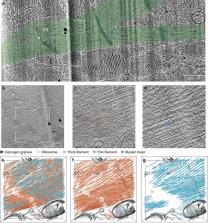
          Sarcomeres, the basic contractile units of striated muscle, produce the forces driving muscular contraction through cross-bridge interactions between actin-containing thin filaments and myosin II-based thick filaments. Until now, direct visualization of the molecular architecture underlying sarcomere contractility has remained elusive. Here, we use in situ cryo-electron tomography to unveil sarcomere contraction in frozen-hydrated neonatal rat cardiomyocytes. We show that the hexagonal lattice of the thick filaments is already established at the neonatal stage, with an excess of thin filaments outside the trigonal positions. Structural assessment of actin polarity by subtomogram averaging reveals that thin filaments in the fully activated state form overlapping arrays of opposite polarity in the center of the sarcomere. Our approach provides direct evidence for thin filament sliding during muscle contraction and may serve as a basis for structural understanding of thin filament activation and actomyosin interactions inside unperturbed cellular environments.

          Abstract

          Sarcomeres, the building blocks of striated muscles, comprise ordered actomyosin arrays involved in force production. Here, the authors visualize sarcomere organization in neonatal cardiomyocytes with in situ cryo-electron tomography, revealing a reduced order of the thin filaments, their sliding and functional states enabling contraction.

          Related collections

          Most cited references73

          • Record: found
          • Abstract: found
          • Article: not found

          UCSF Chimera--a visualization system for exploratory research and analysis.

          The design, implementation, and capabilities of an extensible visualization system, UCSF Chimera, are discussed. Chimera is segmented into a core that provides basic services and visualization, and extensions that provide most higher level functionality. This architecture ensures that the extension mechanism satisfies the demands of outside developers who wish to incorporate new features. Two unusual extensions are presented: Multiscale, which adds the ability to visualize large-scale molecular assemblies such as viral coats, and Collaboratory, which allows researchers to share a Chimera session interactively despite being at separate locales. Other extensions include Multalign Viewer, for showing multiple sequence alignments and associated structures; ViewDock, for screening docked ligand orientations; Movie, for replaying molecular dynamics trajectories; and Volume Viewer, for display and analysis of volumetric data. A discussion of the usage of Chimera in real-world situations is given, along with anticipated future directions. Chimera includes full user documentation, is free to academic and nonprofit users, and is available for Microsoft Windows, Linux, Apple Mac OS X, SGI IRIX, and HP Tru64 Unix from http://www.cgl.ucsf.edu/chimera/. Copyright 2004 Wiley Periodicals, Inc.
            Bookmark
            • Record: found
            • Abstract: found
            • Article: not found

            MotionCor2: anisotropic correction of beam-induced motion for improved cryo-electron microscopy

            MotionCor2 software corrects for beam-induced sample motion, improving the resolution of cryo-EM reconstructions.
              Bookmark
              • Record: found
              • Abstract: found
              • Article: not found

              CTFFIND4: Fast and accurate defocus estimation from electron micrographs.

              CTFFIND is a widely-used program for the estimation of objective lens defocus parameters from transmission electron micrographs. Defocus parameters are estimated by fitting a model of the microscope's contrast transfer function (CTF) to an image's amplitude spectrum. Here we describe modifications to the algorithm which make it significantly faster and more suitable for use with images collected using modern technologies such as dose fractionation and phase plates. We show that this new version preserves the accuracy of the original algorithm while allowing for higher throughput. We also describe a measure of the quality of the fit as a function of spatial frequency and suggest this can be used to define the highest resolution at which CTF oscillations were successfully modeled.
                Bookmark

                Author and article information

                Contributors
                jasnin@biochem.mpg.de
                Journal
                Nat Commun
                Nat Commun
                Nature Communications
                Nature Publishing Group UK (London )
                2041-1723
                2 July 2021
                2 July 2021
                2021
                : 12
                : 4086
                Affiliations
                [1 ]GRID grid.418615.f, ISNI 0000 0004 0491 845X, Department of Cellular and Molecular Biophysics, , Max Planck Institute of Biochemistry, ; Martinsried, Germany
                [2 ]GRID grid.418615.f, ISNI 0000 0004 0491 845X, Department of Molecular Structural Biology, , Max Planck Institute of Biochemistry, ; Martinsried, Germany
                [3 ]GRID grid.418615.f, ISNI 0000 0004 0491 845X, Department of Molecular Medicine, , Max Planck Institute of Biochemistry, ; Martinsried, Germany
                Author information
                http://orcid.org/0000-0003-3050-7163
                http://orcid.org/0000-0002-6106-4847
                http://orcid.org/0000-0002-6402-8315
                http://orcid.org/0000-0003-1726-4566
                Article
                24049
                10.1038/s41467-021-24049-0
                8253822
                34215727
                87b5515f-57e2-48d9-b553-8415c9e8a596
                © The Author(s) 2021

                Open Access This article is licensed under a Creative Commons Attribution 4.0 International License, which permits use, sharing, adaptation, distribution and reproduction in any medium or format, as long as you give appropriate credit to the original author(s) and the source, provide a link to the Creative Commons license, and indicate if changes were made. The images or other third party material in this article are included in the article’s Creative Commons license, unless indicated otherwise in a credit line to the material. If material is not included in the article’s Creative Commons license and your intended use is not permitted by statutory regulation or exceeds the permitted use, you will need to obtain permission directly from the copyright holder. To view a copy of this license, visit http://creativecommons.org/licenses/by/4.0/.

                History
                : 24 September 2020
                : 27 May 2021
                Funding
                Funded by: Deutsches Zentrum für Herz-Kreislaufforschung (grant 81Z1600313).
                Categories
                Article
                Custom metadata
                © The Author(s) 2021

                Uncategorized
                actin,myosin,cryoelectron tomography
                Uncategorized
                actin, myosin, cryoelectron tomography

                Comments

                Comment on this article