2
views
0
recommends
+1 Recommend
0 collections
    0
    shares
      • Record: found
      • Abstract: found
      • Article: found
      Is Open Access

      TomoTwin: generalized 3D localization of macromolecules in cryo-electron tomograms with structural data mining

      research-article

      Read this article at

      Bookmark
          There is no author summary for this article yet. Authors can add summaries to their articles on ScienceOpen to make them more accessible to a non-specialist audience.

          Abstract

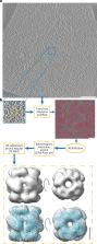
          Cryogenic-electron tomography enables the visualization of cellular environments in extreme detail, however, tools to analyze the full amount of information contained within these densely packed volumes are still needed. Detailed analysis of macromolecules through subtomogram averaging requires particles to first be localized within the tomogram volume, a task complicated by several factors including a low signal to noise ratio and crowding of the cellular space. Available methods for this task suffer either from being error prone or requiring manual annotation of training data. To assist in this crucial particle picking step, we present TomoTwin: an open source general picking model for cryogenic-electron tomograms based on deep metric learning. By embedding tomograms in an information-rich, high-dimensional space that separates macromolecules according to their three-dimensional structure, TomoTwin allows users to identify proteins in tomograms de novo without manually creating training data or retraining the network to locate new proteins.

          Abstract

          TomoTwin is a deep metric learning-based particle picking method for cryo-electron tomograms. TomoTwin obviates the need for annotating training data and retraining a picking model for each protein.

          Related collections

          Most cited references60

          • Record: found
          • Abstract: found
          • Article: found
          Is Open Access

          New tools for automated high-resolution cryo-EM structure determination in RELION-3

          Here, we describe the third major release of RELION. CPU-based vector acceleration has been added in addition to GPU support, which provides flexibility in use of resources and avoids memory limitations. Reference-free autopicking with Laplacian-of-Gaussian filtering and execution of jobs from python allows non-interactive processing during acquisition, including 2D-classification, de novo model generation and 3D-classification. Per-particle refinement of CTF parameters and correction of estimated beam tilt provides higher resolution reconstructions when particles are at different heights in the ice, and/or coma-free alignment has not been optimal. Ewald sphere curvature correction improves resolution for large particles. We illustrate these developments with publicly available data sets: together with a Bayesian approach to beam-induced motion correction it leads to resolution improvements of 0.2–0.7 Å compared to previous RELION versions.
            Bookmark
            • Record: found
            • Abstract: found
            • Article: not found

            Automated electron microscope tomography using robust prediction of specimen movements.

            A new method was developed to acquire images automatically at a series of specimen tilts, as required for tomographic reconstruction. The method uses changes in specimen position at previous tilt angles to predict the position at the current tilt angle. Actual measurement of the position or focus is skipped if the statistical error of the prediction is low enough. This method allows a tilt series to be acquired rapidly when conditions are good but falls back toward the traditional approach of taking focusing and tracking images when necessary. The method has been implemented in a program, SerialEM, that provides an efficient environment for data acquisition. This program includes control of an energy filter as well as a low-dose imaging mode, in which tracking and focusing occur away from the area of interest. The program can automatically acquire a montage of overlapping frames, allowing tomography of areas larger than the field of the CCD camera. It also includes tools for navigating between specimen positions and finding regions of interest.
              Bookmark
              • Record: found
              • Abstract: found
              • Article: not found

              Computer visualization of three-dimensional image data using IMOD.

              We have developed a computer software package, IMOD, as a tool for analyzing and viewing three-dimensional biological image data. IMOD is useful for studying and modeling data from tomographic, serial section, and optical section reconstructions. The software allows image data to be visualized by several different methods. Models of the image data can be visualized by volume or contour surface rendering and can yield quantitative information.
                Bookmark

                Author and article information

                Contributors
                stefan.raunser@mpi-dortmund.mpg.de
                Journal
                Nat Methods
                Nat Methods
                Nature Methods
                Nature Publishing Group US (New York )
                1548-7091
                1548-7105
                15 May 2023
                15 May 2023
                2023
                : 20
                : 6
                : 871-880
                Affiliations
                GRID grid.418441.c, ISNI 0000 0004 0491 3333, Department of Structural Biochemistry, , Max Planck Institute of Molecular Physiology, ; Dortmund, Germany
                Author information
                http://orcid.org/0000-0002-1550-8275
                http://orcid.org/0000-0003-0191-6419
                http://orcid.org/0000-0002-4496-7489
                http://orcid.org/0000-0001-9373-3016
                Article
                1878
                10.1038/s41592-023-01878-z
                10250198
                37188953
                010e98d1-3463-44cb-a9f9-4f8a74bc2acc
                © The Author(s) 2023

                Open Access This article is licensed under a Creative Commons Attribution 4.0 International License, which permits use, sharing, adaptation, distribution and reproduction in any medium or format, as long as you give appropriate credit to the original author(s) and the source, provide a link to the Creative Commons license, and indicate if changes were made. The images or other third party material in this article are included in the article’s Creative Commons license, unless indicated otherwise in a credit line to the material. If material is not included in the article’s Creative Commons license and your intended use is not permitted by statutory regulation or exceeds the permitted use, you will need to obtain permission directly from the copyright holder. To view a copy of this license, visit http://creativecommons.org/licenses/by/4.0/.

                History
                : 24 June 2022
                : 12 April 2023
                Funding
                Funded by: FundRef https://doi.org/10.13039/501100004189, Max-Planck-Gesellschaft (Max Planck Society);
                Categories
                Article
                Custom metadata
                © Springer Nature America, Inc. 2023

                Life sciences
                image processing,software,cryoelectron tomography
                Life sciences
                image processing, software, cryoelectron tomography

                Comments

                Comment on this article