6
views
0
recommends
+1 Recommend
0 collections
    0
    shares
      • Record: found
      • Abstract: found
      • Article: found
      Is Open Access

      Coexistence of condyloma acuminatum and extramammary Paget’s disease on penis and scrotum: A rare case report

      research-article

      Read this article at

      Bookmark
          There is no author summary for this article yet. Authors can add summaries to their articles on ScienceOpen to make them more accessible to a non-specialist audience.

          Rationale:

          Extramammary Paget’s disease (EMPD) is a rare skin cancer that commonly occurs in sites rich in apocrine glands, such as perineum, vulva, axilla, scrotum, and penis. On the other hand, condyloma acuminatum (CA; also referred to as anogenital warts) is a common benign neoplasm caused by human papillomavirus. Few cases of coexistent EMPD and CA have been reported because of the rarity of the condition.

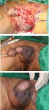
          Patient concerns and diagnosis:

          A 72-year-old man with a genital mass, which appeared to be composed of multiple papillomatous masses, was referred for surgical resection. The lesion was first noticed 6 months previously and grew rapidly. CO 2 ablative laser therapy was performed twice at a primary clinic, but the mass recurred.

          Intervention and outcomes:

          Excisional biopsy revealed the presence of coexistent EMPD and CA. Additional wide excision was performed, and postoperative biopsy confirmed no residual tumor. Two years after surgery, no recurrence had occurred.

          Lessons:

          CA can co-occur with several types of skin malignancies, and a skin malignancy coexisting with CA is difficult to diagnose visually. Therefore, even if a skin lesion in the genital region is considered to be CA, we recommend punch biopsy before treatment because it can benefit prognosiss.

          Related collections

          Most cited references16

          • Record: found
          • Abstract: found
          • Article: not found

          Mammary and extramammary Paget's disease.

          Mammary and extramammary Paget's disease are uncommon intraepithelial adenocarcinomas. Both conditions have similar clinical features, which mimic inflammatory and infective diseases. Histological diagnostic confusion can arise between Paget's disease and other neoplastic conditions affecting the skin, with the most common differential diagnoses being malignant melanoma and atypical squamous disease. The glandular differentiation of both mammary Paget's disease and extramammary Paget's disease is indicated by morphological appearances, the presence of intracellular mucin in many cases, and positive immunohistochemical staining for glandular cytokeratins, epithelial membrane antigen, and carcinoembryonic antigen. This article provides an overview of mammary and extramammary Paget's disease and discusses recent evidence regarding the cell of origin. The concepts of primary and secondary Paget's disease are presented and the differential diagnosis is discussed with reference to immunohistochemical markers that might be of diagnostic value.
            Bookmark
            • Record: found
            • Abstract: found
            • Article: not found

            The role of immunohistochemistry in discriminating primary from secondary extramammary Paget disease.

            Extramammary Paget disease (EMPD) is categorized into 2 groups: primary EMPD or EMPD secondary to underlying malignancy. Primary EMPD has a better prognosis, and the ability to distinguish between the 2 subsets has clinical relevance. Recent studies have suggested that immunostains, including cytokeratin (CK) 7, CK20, and BRST-2, distinguish between the 2 groups. We analyzed a large series of EMPD with an expanded immunohistochemical panel to assess its value in distinguishing primary from secondary disease.
              Bookmark
              • Record: found
              • Abstract: found
              • Article: not found

              Extramammary Paget's disease of scrotum--report of 25 cases and literature review.

              We evaluate the clinical manifestations, management, and prognostic characteristics of scrotal extramammary Paget's disease (EMPD). The study comprised 25 patients with scrotal EMPD at our institute from January 1982 to February 2005, with all available clinical and pathological data reviewed. Of these 25 patients, 1 received radiotherapy and 24 received local wide excisions. In 24 operated patients, 7 had local recurrence and/or metastasis of groin lymph node. Five of the 7 with recurrence had a positive surgical margin postoperatively and they received a second local extensive excision. One of the 7 with recurrence and metastasis of the groin lymph node had a second local extensive excision with groin lymphadenectomy, and the last one who only had metastasis of the groin lymph node had a groin lymphadenectomy. Four of 13 patients with dermal invasion by Paget's cell had metastasis. None of the other 12 patients without dermal invasion had metastasis. However, there was no statistical metastasis rate difference (P = 0.096) between the patients with dermal invasion by Paget's cell and without. There was no statistical difference (P = 0.947) in mean delay time from onset of symptoms to diagnosis between the 2 groups either. The follow-up duration varies from 17 to 243 months (mean 119 + 86.2 months). One patient with stage D died of EMPD of the scrotum. We found that EMPD of the scrotum is usually a slow progressive disease, mainly seen in elderly patients, and has a good prognosis when there is noninvasive disease. The primary treatment for EMPD of the scrotum is wide surgical excision. The key to decreasing tumor recurrence, however, is a precise, preoperative histological examination to define the range of the lesion. Copyright 2010 Elsevier Inc. All rights reserved.
                Bookmark

                Author and article information

                Contributors
                Journal
                Medicine (Baltimore)
                Medicine (Baltimore)
                MD
                Medicine
                Lippincott Williams & Wilkins (Hagerstown, MD )
                0025-7974
                1536-5964
                11 November 2022
                11 November 2022
                : 101
                : 45
                : e31754
                Affiliations
                [a ] Department of Plastic and Reconstructive Surgery, College of Medicine, Dongguk University, Gyeongju-si, Republic of Korea
                [b ] Pathology, College of Medicine, Dongguk University, Gyeongju-si, Republic of Korea
                [c ] Department of Plastic and Reconstructive Surgery, Institute for Human Tissue Restoration, Severance Hospital, Yonsei University College of Medicine, Seoul, Republic of Korea.
                Author notes
                * Correspondence: Gyu Yong Jung, Department of Plastic and Reconstructive Surgery, College of Medicine, Dongguk University, 87 Dongdae-ro, Gyeongju 38067, Republic of Korea (e-mail: jnjbeauty@ 123456naver.com ).
                Author information
                https://orcid.org/0000-0002-7098-6958
                https://orcid.org/0000-0002-0653-9766
                Article
                00059
                10.1097/MD.0000000000031754
                9666121
                36397375
                7ae25ead-98aa-45b0-ae70-b01dae7a80a6
                Copyright © 2022 the Author(s). Published by Wolters Kluwer Health, Inc.

                This is an open access article distributed under the Creative Commons Attribution License 4.0 (CCBY), which permits unrestricted use, distribution, and reproduction in any medium, provided the original work is properly cited.

                History
                : 03 August 2022
                : 21 October 2022
                Categories
                4000
                Research Article
                Clinical Case Report
                Custom metadata
                TRUE

                case report,condyloma acuminatum,extramammary paget disease,human papillomavirus

                Comments

                Comment on this article