75
views
0
recommends
+1 Recommend
0 collections
    0
    shares
      • Record: found
      • Abstract: found
      • Article: found
      Is Open Access

      Heparanase mediates renal dysfunction during early sepsis in mice

      research-article

      Read this article at

      Bookmark
          There is no author summary for this article yet. Authors can add summaries to their articles on ScienceOpen to make them more accessible to a non-specialist audience.

          Abstract

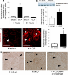
          Heparanase, a heparan sulfate-specific glucuronidase, mediates the onset of pulmonary neutrophil adhesion and inflammatory lung injury during early sepsis. We hypothesized that glomerular heparanase is similarly activated during sepsis and contributes to septic acute kidney injury (AKI). We induced polymicrobial sepsis in mice using cecal ligation and puncture (CLP) in the presence or absence of competitive heparanase inhibitors (heparin or nonanticoagulant N-desulfated re- N-acetylated heparin [NAH]). Four hours after surgery, we collected serum and urine for measurement of renal function and systemic inflammation, invasively determined systemic hemodynamics, harvested kidneys for histology/protein/mRNA, and/or measured glomerular filtration by inulin clearance. CLP-treated mice demonstrated early activation of glomerular heparanase with coincident loss of glomerular filtration, as indicated by a >twofold increase in blood urea nitrogen (BUN) and a >50% decrease in inulin clearance ( P < 0.05) in comparison to sham mice. Administration of heparanase inhibitors 2 h prior to CLP attenuated sepsis-induced loss of glomerular filtration rate, demonstrating that heparanase activation contributes to early septic renal dysfunction. Glomerular heparanase activation was not associated with renal neutrophil influx or altered vascular permeability, in marked contrast to previously described effects of pulmonary heparanase on neutrophilic lung injury during sepsis. CLP induction of renal inflammatory gene (IL-6, TNF-α, IL-1β) expression was attenuated by NAH pretreatment. While serum inflammatory indices (KC, IL-6, TNF-α, IL-1β) were not impacted by NAH pretreatment, heparanase inhibition attenuated the CLP-induced increase in serum IL-10. These findings demonstrate that glomerular heparanase is active during sepsis and contributes to septic renal dysfunction via mechanisms disparate from heparanase-mediated lung injury.

          Related collections

          Most cited references29

          • Record: found
          • Abstract: found
          • Article: not found

          Sepsis and acute kidney injury.

          Sepsis is a severe and dysregulated inflammatory response to infection characterized by end-organ dysfunction distant from the primary site of infection. Development of acute kidney injury (AKI) during sepsis increases patient morbidity, predicts higher mortality, has a significant effect on multiple organ functions, is associated with an increased length of stay in the intensive care unit, and hence consumes considerable healthcare resources. When compared with AKI of nonseptic origin, septic AKI is characterized by a distinct pathophysiology and therefore requires a different approach. Despite impressive advances in several fields of medicine, the pathophysiology, diagnostic procedures, and appropriate therapeutic interventions in sepsis are still highly debatable. Numerous immunomodulatory agents showing promise in preclinical studies fail to reduce the overwhelmingly high mortality rate of sepsis and provoke AKI when compared with other critically ill patients. Major impediments to progress in understanding, early diagnosis, and application of appropriate therapeutic modalities in sepsis-induced AKI include limited histopathologic information, few animal models that closely mimic human sepsis, and a relative shortage of specific diagnostic tools. Here we discuss the most recent advances in understanding the fundamental mechanisms of sepsis-induced AKI, characteristics of relevant animal models available, and potential therapies.
            Bookmark
            • Record: found
            • Abstract: found
            • Article: not found

            Mechanisms of cardiac and renal dysfunction in patients dying of sepsis.

            The mechanistic basis for cardiac and renal dysfunction in sepsis is unknown. In particular, the degree and type of cell death is undefined. To evaluate the degree of sepsis-induced cardiomyocyte and renal tubular cell injury and death. Light and electron microscopy and immunohistochemical staining for markers of cellular injury and stress, including connexin-43 and kidney-injury-molecule-1 (Kim-1), were used in this study. Rapid postmortem cardiac and renal harvest was performed in 44 septic patients. Control hearts were obtained from 12 transplant and 13 brain-dead patients. Control kidneys were obtained from 20 trauma patients and eight patients with cancer. Immunohistochemistry demonstrated low levels of apoptotic cardiomyocytes (<1-2 cells per thousand) in septic and control subjects and revealed redistribution of connexin-43 to lateral membranes in sepsis (P < 0.020). Electron microscopy showed hydropic mitochondria only in septic specimens, whereas mitochondrial membrane injury and autophagolysosomes were present equally in control and septic specimens. Control kidneys appeared relatively normal by light microscopy; 3 of 20 specimens showed focal injury in approximately 1% of renal cortical tubules. Conversely, focal acute tubular injury was present in 78% of septic kidneys, occurring in 10.3 ± 9.5% and 32.3 ± 17.8% of corticomedullary-junction tubules by conventional light microscopy and Kim-1 immunostains, respectively (P < 0.01). Electron microscopy revealed increased tubular injury in sepsis, including hydropic mitochondria and increased autophagosomes. Cell death is rare in sepsis-induced cardiac dysfunction, but cardiomyocyte injury occurs. Renal tubular injury is common in sepsis but presents focally; most renal tubular cells appear normal. The degree of cell injury and death does not account for severity of sepsis-induced organ dysfunction.
              Bookmark
              • Record: found
              • Abstract: found
              • Article: not found

              Heparan sulfate proteoglycan is a mechanosensor on endothelial cells.

              The objective of this study was to test whether a glycosaminoglycan component of the surface glycocalyx layer is a fluid shear stress sensor on endothelial cells (ECs). Because enhanced nitric oxide (NO) production in response to fluid shear stress is a characteristic and physiologically important response of ECs, we evaluated NOx (NO2- and NO3-) production in response to fluid shear stress after enzymatic removal of heparan sulfate, the dominant glycosaminoglycan of the EC glycocalyx, from cultured ECs. The significant NOx production induced by steady shear stress (20 dyne/cm2) was inhibited completely by pretreatment with 15 mU/mL heparinase III (E.C.4.2.2.8) for 2 hours. Oscillatory shear stress (10+/-15 dyne/cm2) induced an even greater NOx production than steady shear stress that was completely inhibited by pretreatment with heparinase III. Addition of bradykinin (BK) induced significant NOx production that was not inhibited by heparinase pretreatment, demonstrating that the cells were still able to produce abundant NO after heparinase treatment. Fluorescent imaging with a heparan sulfate antibody revealed that heparinase III treatments removed a substantial fraction of the heparan sulfate bound to the surfaces of ECs. In summary, these experiments demonstrate that a heparan sulfate component of the EC glycocalyx participates in mechanosensing that mediates NO production in response to shear stress. The full text of this article is available online at http://www.circresaha.org.
                Bookmark

                Author and article information

                Journal
                Physiol Rep
                Physiol Rep
                phy2
                Physiological Reports
                Blackwell Publishing Ltd
                2051-817X
                2051-817X
                November 2013
                11 November 2013
                : 1
                : 6
                : e00153
                Affiliations
                [1 ]Program in Translational Lung Research, Division of Pulmonary Sciences and Critical Care Medicine, University of Colorado School of Medicine Aurora, Colorado
                [2 ]Division of Renal Diseases and Hypertension, University of Colorado School of Medicine Aurora, Colorado
                [3 ]Denver Health Medical Center Denver, Colorado
                Author notes
                Eric Schmidt, Research Complex 2, Mail Stop C272, 12700 E. 19th Avenue, Aurora, CO 80045., Tel: 303-724-6106, Fax: 303-724-6042, E-mail: eric.schmidt@ 123456ucdenver.edu

                Funding Information E. P. S. is currently receiving a grant (K08 HL105538) from the NIH/NHLBI; S. F. is currently receiving a grant (R01 HL095363) from the NIH/NHLBI; R. M. T. is currently receiving a grant (R01 ES016285) from the NIH/NHLBI.

                Article
                10.1002/phy2.153
                3871468
                24400155
                796addf9-23b1-4a5d-a230-fcdddcac0437
                © 2013 The Authors. Physiological Reports published by Wiley Periodicals, Inc. on behalf of the American Physiological Society and The Physiological Society

                Re-use of this article is permitted in accordance with the Creative Commons Deed, Attribution 2.5, which does not permit commercial exploitation.

                History
                : 30 July 2013
                : 26 September 2013
                : 16 October 2013
                Categories
                Original Research

                acute kidney injury,heparan sulfate,heparanase,heparin,mice,sepsis

                Comments

                Comment on this article