Inviting an author to review:
Find an author and click ‘Invite to review selected article’ near their name.
Search for authorsSearch for similar articles
12
views
0
recommends
+1 Recommend
0 collections
    0
    shares
      • Record: found
      • Abstract: found
      • Article: found
      Is Open Access

      A case report describing the successful separation of ischiopagus tetrapus conjoined twins in Vietnam

      case-report

      Read this article at

      Bookmark
          There is no author summary for this article yet. Authors can add summaries to their articles on ScienceOpen to make them more accessible to a non-specialist audience.

          Abstract

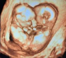
          Ischiopagus conjoined twinning is a rare congenital defect. The surgical separation of conjoined twins is difficult because of the complex anatomy and physiology. Careful preoperative assessment, planning, and effective surgical teams are critically important for achieving a good outcome. We describe the successful separation of ischiopagus tetrapus conjoined twins as a representative case demonstrating the growth of pediatric surgery in southern Vietnam.

          Related collections

          Most cited references12

          • Record: found
          • Abstract: found
          • Article: not found

          Surgical experience with thirteen conjoined twins.

          Conjoined twins occur in approximately one in 50,000 or so births, and most do not survive. The authors report herein their experience with 13 conjoined twins over the last 30 years, involving those of the following forms: thoracopagus (4 cases), omphalopagus (1 case), ischiopagus (4 cases), pygopagus (1 case), craniopagus (1 case), and incomplete or parasitic varieties (2 cases). The various diagnostic and imaging studies used are described in detail for each form of twinning. Separation is best delayed until such infants are relatively mature (i.e., 6-12 months of age). Operative survival was 50% in those operated on in the neonatal period, but 90% in those over 4 months of age. Ten separations were attempted in 13 sets of twins, with 16 operative survivors. Significantly, up to 10 years after surgery, there were six late deaths due to serious associated congenital anomalies, predominantly cardiac. Improved recent survival is probably the result of the availability of more accurate imaging studies and better anesthetic and operative techniques, with great emphasis on performing immediate reconstruction whenever possible. Use of skin expanders and prosthetic mesh has facilitated wound closure. In the future, ex vivo cardiac reconstruction and autotransplantation may permit separation of twins with complicated conjoined hearts.
            Bookmark
            • Record: found
            • Abstract: found
            • Article: not found

            Conjoined twins: From conception to separation, a review.

            Conjoined twins are suggested to result from aberrant embryogenesis. The two main theories proposed to explain the phenomena are fission and fusion. The incidence rate is 1 in 50,000 births; however, since about 60% of the cases are stillborn, the true incidence is approximated at 1 in 200,000. There is a higher predisposition towards female than male gender with a ratio of 3:1. Conjoined twins are classified based upon the site of attachment. The extent of organ sharing, especially the heart, determines the possibility and prognosis of a separation procedure. Meticulous preoperative evaluation, planning, and preparedness of the team are crucial for a successful separation. Separation of conjoined twins poses several technical, legal, and ethical issues. Clin. Anat. 30:385-396, 2017. © 2017 Wiley Periodicals, Inc.
              Bookmark
              • Record: found
              • Abstract: found
              • Article: found
              Is Open Access

              Conjoined twins – role of imaging and recent advances

              Introduction Conjoined twins are identical twins with fused bodies, joined in utero. They are rare complications of monochorionic twinning. The purpose of this study is to describe the various types of conjoined twins, the role of imaging and recent advances aiding in their management. Material and methods This was a twin institutional study involving 3 cases of conjoined twins diagnosed over a period of 6 years from 2010 to 2015. All the 3 cases were identified antenatally by ultrasound. Only one case was further evaluated by MRI. Results Three cases of conjoined twins (cephalopagus, thoracopagus and omphalopagus) were accurately diagnosed on antenatal ultrasound. After detailed counseling of the parents and obtaining written consent, all the three cases of pregnancy were terminated. Delivery of the viable conjoined twins was achieved without any complications to the mothers, and all the three conjoined twins died after a few minutes. Conclusion Ultrasound enables an early and accurate diagnosis of conjoined twins, which is vital for obstetric management. MRI is reserved for better tissue characterization. Termination of pregnancy when opted, should be done at an early stage as later stages are fraught with problems. Recent advances, such as 3D printing, may aid in surgical pre-planning, thereby enabling successful surgical separation of conjoined twins.
                Bookmark

                Author and article information

                Contributors
                Journal
                Radiol Case Rep
                Radiol Case Rep
                Radiology Case Reports
                Elsevier
                1930-0433
                15 July 2021
                September 2021
                15 July 2021
                : 16
                : 9
                : 2658-2662
                Affiliations
                [a ]Department of General Surgery, Ho Chi Minh City Children's Hospital, Ho Chi Minh City, Vietnam
                [b ]Department of Radiology, Hanoi Medical University, Ha Noi, Viet Nam
                [c ]Department of Radiology, Pham Ngoc Thach University of Medicine, Ho Chi Minh City, Viet Nam
                [d ]Department of Radiology, Children's Hospital 2, Ho Chi Minh City, Viet Nam
                [e ]Department of Neonatal Intensive Care, Ho Chi Minh City Children's Hospital, Ho Chi Minh City, Vietnam
                [f ]Department of Anesthesiology, Ho Chi Minh City Children's Hospital, Ho Chi Minh City, Vietnam
                Author notes
                [1]

                These authors contributed equally to this article as co-first authors.

                Article
                S1930-0433(21)00424-6
                10.1016/j.radcr.2021.06.048
                8319480
                34345326
                783f1780-3e55-40c3-8d5e-366d8c5c1073
                © 2021 The Authors. Published by Elsevier Inc. on behalf of University of Washington.

                This is an open access article under the CC BY-NC-ND license (http://creativecommons.org/licenses/by-nc-nd/4.0/).

                History
                : 12 June 2021
                : 14 June 2021
                : 16 June 2021
                Categories
                Case Report

                ischiopagus tetrapus,conjoined twins,twin separation surgery

                Comments

                Comment on this article

                scite_
                0
                0
                0
                0
                Smart Citations
                0
                0
                0
                0
                Citing PublicationsSupportingMentioningContrasting
                View Citations

                See how this article has been cited at scite.ai

                scite shows how a scientific paper has been cited by providing the context of the citation, a classification describing whether it supports, mentions, or contrasts the cited claim, and a label indicating in which section the citation was made.

                Similar content390

                Cited by1

                Most referenced authors133