Inviting an author to review:
Find an author and click ‘Invite to review selected article’ near their name.
Search for authorsSearch for similar articles
24
views
0
recommends
+1 Recommend
0 collections
    0
    shares
      • Record: found
      • Abstract: found
      • Article: found
      Is Open Access

      Surgical Correction of Ischiopagus Tripus Conjoined Twins with Fused Pelvis: Enhancing Quality of Life through Orthopedic Intervention

      research-article

      Read this article at

      Bookmark
          There is no author summary for this article yet. Authors can add summaries to their articles on ScienceOpen to make them more accessible to a non-specialist audience.

          Abstract

          Patient: Male, 3-year-old

          Final Diagnosis: Ischiopagus tripus conjoined twins after surgical correction

          Symptoms: Fused body in pelvis reion

          Clinical Procedure: Amputation • osteotomy

          Specialty: Orthopedics and Traumatology

          Objective:

          Congenital defects/diseases

          Background:

          The rarity of ischiopagus tripus conjoined twins complicates the surgical separation, owing to the lack of cases and high complexity. We aim to report our experience in performing orthopedic correction for ischiopagus tripus twins.

          Case Report:

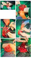
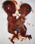
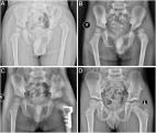
          A pair of 3-year-old conjoined boys presented with a fused body at the pelvis region and only 1 umbilicus. There were 2 legs separated by shared genitalia and an anus at the midline, and 1 fused leg, which could be felt and moved by both of the patients. The twins also shared internal organs of the bladder, intestine, and rectum, as visualized through angiography computerized tomography scan. After several team discussions with the institutional review board, the hospital ethics committee, and both parents, it was agreed to perform disarticulation of the fused third limb, followed by correction of the trunk alignment by pelvic closed wedge osteotomy and internal fixation. We successfully reconstructed the pelvis using locking plates and additional 3.5-mm cortical screws and 1.2-mm stainless steel wire.

          Conclusions:

          This report describes the presentation and surgical management of a case of ischiopagus tripus conjoined twins. It highlights the challenges involved in surgery and the importance of investigating these infants for other congenital abnormalities. Although surgical approaches for different sets of twins should be individually tailored, interventions aimed to provide optimal outcomes should consider ethical issues and parental/patient expectations. Even in situations in which the twins are inseparable, there is still room for surgical correction to be performed.

          Related collections

          Most cited references17

          • Record: found
          • Abstract: found
          • Article: not found

          Conjoined twins: From conception to separation, a review.

          Conjoined twins are suggested to result from aberrant embryogenesis. The two main theories proposed to explain the phenomena are fission and fusion. The incidence rate is 1 in 50,000 births; however, since about 60% of the cases are stillborn, the true incidence is approximated at 1 in 200,000. There is a higher predisposition towards female than male gender with a ratio of 3:1. Conjoined twins are classified based upon the site of attachment. The extent of organ sharing, especially the heart, determines the possibility and prognosis of a separation procedure. Meticulous preoperative evaluation, planning, and preparedness of the team are crucial for a successful separation. Separation of conjoined twins poses several technical, legal, and ethical issues. Clin. Anat. 30:385-396, 2017. © 2017 Wiley Periodicals, Inc.
            Bookmark
            • Record: found
            • Abstract: found
            • Article: found
            Is Open Access

            Conjoined twins – role of imaging and recent advances

            Introduction Conjoined twins are identical twins with fused bodies, joined in utero. They are rare complications of monochorionic twinning. The purpose of this study is to describe the various types of conjoined twins, the role of imaging and recent advances aiding in their management. Material and methods This was a twin institutional study involving 3 cases of conjoined twins diagnosed over a period of 6 years from 2010 to 2015. All the 3 cases were identified antenatally by ultrasound. Only one case was further evaluated by MRI. Results Three cases of conjoined twins (cephalopagus, thoracopagus and omphalopagus) were accurately diagnosed on antenatal ultrasound. After detailed counseling of the parents and obtaining written consent, all the three cases of pregnancy were terminated. Delivery of the viable conjoined twins was achieved without any complications to the mothers, and all the three conjoined twins died after a few minutes. Conclusion Ultrasound enables an early and accurate diagnosis of conjoined twins, which is vital for obstetric management. MRI is reserved for better tissue characterization. Termination of pregnancy when opted, should be done at an early stage as later stages are fraught with problems. Recent advances, such as 3D printing, may aid in surgical pre-planning, thereby enabling successful surgical separation of conjoined twins.
              Bookmark
              • Record: found
              • Abstract: found
              • Article: found
              Is Open Access

              Pelvic osteotomies in hip dysplasia: why, when and how?

              Acetabular dysplasia is a significant problem in the spectrum of developmental dysplasia of hip. In a younger child, positioning the femoral head into the acetabulum helps in reciprocal remodeling of the acetabulum and correction of dysplasia. In an older child, the remodeling potential is limited and often the acetabular dysplasia needs surgical intervention in the form of a pelvic osteotomy. Thus, pelvic osteotomy forms an integral part of surgical management of hip dysplasia. The ultimate goal of these osteotomies is to preclude or postpone the development of osteoarthritis and add more years of life to the native hip. Pelvic osteotomies play a pivotal role in normalizing hip morphology. The choice of pelvic osteotomy depends on the age of a child, the type of dysplasia and the status of the tri-radiate cartilage. Several types of re-directional and reshaping pelvic osteotomies have been described in the literature to improve the stability and restore the anatomy and biomechanics of the dysplastic hip. This article attempts to review the current indications for various pelvic osteotomies with a brief description of their techniques along with the outcomes and complications published thus far. Besides, the guidelines to choose the right pelvic osteotomy are also provided.
                Bookmark

                Author and article information

                Journal
                Am J Case Rep
                Am J Case Rep
                amjcaserep
                The American Journal of Case Reports
                International Scientific Literature, Inc.
                1941-5923
                2024
                12 May 2024
                : 25
                : e942126-1-e942126-8
                Affiliations
                [1 ]Division of Pediatric Orthopedics, Department of Orthopedics and Traumatology, Hasan Sadikin General Hospital/Faculty of Medicine, University of Padjadjaran, Bandung, West Java, Indonesia
                [2 ]Faculty of Medicine, University of Sriwijaya, Palembang, South Sumatra, Indonesia
                [3 ]Division of Orthopedics and Traumatology, Department of Surgery, Sardjito General Hospital/Faculty of Medicine, Public Health and Nursing, Universitas Gadjah Mada, Yogyakarta, Indonesia
                Author notes
                Corresponding Author: Hilmi Muhammad, e-mail: hilmimuhammadortho@ 123456gmail.com

                Authors’ Contribution:

                [A]

                Study Design

                [B]

                Data Collection

                [C]

                Statistical Analysis

                [D]

                Data Interpretation

                [E]

                Manuscript Preparation

                [F]

                Literature Search

                [G]

                Funds Collection

                Financial support: None declared

                Conflict of interest: None declared

                Author information
                https://orcid.org/0000-0001-5112-0916
                Article
                942126
                10.12659/AJCR.942126
                11099550
                38734882
                530edee7-21c9-45b9-aa15-cf3692e1a778
                © Am J Case Rep, 2024

                This work is licensed under Creative Common Attribution-NonCommercial-NoDerivatives 4.0 International ( CC BY-NC-ND 4.0)

                History
                : 10 August 2023
                : 20 March 2024
                : 27 March 2024
                Categories
                Articles

                twins, conjoined,orthopedic procedures,pediatrics
                twins, conjoined, orthopedic procedures, pediatrics

                Comments

                Comment on this article

                scite_
                0
                0
                0
                0
                Smart Citations
                0
                0
                0
                0
                Citing PublicationsSupportingMentioningContrasting
                View Citations

                See how this article has been cited at scite.ai

                scite shows how a scientific paper has been cited by providing the context of the citation, a classification describing whether it supports, mentions, or contrasts the cited claim, and a label indicating in which section the citation was made.

                Similar content226

                Most referenced authors103