11
views
0
recommends
+1 Recommend
0 collections
    0
    shares
      • Record: found
      • Abstract: found
      • Article: found
      Is Open Access

      Overexpressed pseudogenes, DUXAP8 and DUXAP9, promote growth of renal cell carcinoma and serve as unfavorable prognostic biomarkers

      research-article

      Read this article at

      Bookmark
          There is no author summary for this article yet. Authors can add summaries to their articles on ScienceOpen to make them more accessible to a non-specialist audience.

          Abstract

          Background: Growing studies have reported that pseudogenes play key roles in multiple human cancers. However, expression and roles of pseudogenes in renal cell carcinoma remains absent.

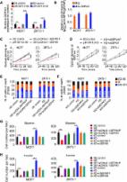
          Results: 31 upregulated and 16 downregulated pseudogenes were screened. Higher expression of DUXAP8 and DUXAP9 indicated poorer prognosis of kidney cancer. 33 and 5 miRNAs were predicted to potentially binding to DUXAP8 and DUXAP9, respectively. miR-29c-3p was identified as the most potential binding miRNAs of DUXAP8 and DUXAP9 based on expression, survival and correlation analyses. 254 target genes of miR-29c-3p were forecast. 47 hub genes with node degree >= 10 were identified. Subsequent analysis for the top 10 hub genes demonstrated that COL1A1 and COL1A2 may be two functional targets of DUXAP8 and DUXAP9. Expression of DUXAP8, DUXAP9, COL1A1 and COL1A2 were significantly increased in cancer samples compared to normal controls while miR-29c-3p expression was decreased. Luciferase reporter assay revealed that miR-29c-3p could directly bind to DUXAP8, DUXAP9, COL1A1 and COL1A2. Functional experiments showed that DUXAP8 and DUXAP9 enhanced but miR-29c-3p weakened growth of renal cell carcinoma.

          Conclusions: In conclusion, upregulated DUXAP8 and DUXAP9 promote growth of renal cell carcinoma and serve as two promising prognostic biomarkers.

          Methods: Dysregulated pseudogenes were obtained by dreamBase and GEPIA. The binding miRNAs of pseudogene and targets of miRNA were predicted using starBase and miRNet. Kaplan-Meier plotter was utilized to perform survival analysis, and Enrichr database was introduced to conduct functional enrichment analysis. Hub genes were identified through STRING and Cytoscape. qRT-PCR, luciferase reporter assay, cell counting assay and colony formation assay were performed to validate in silico analytic results.

          Related collections

          Most cited references37

          • Record: found
          • Abstract: found
          • Article: not found

          Pseudogene PTENP1 functions as a competing endogenous RNA to suppress clear-cell renal cell carcinoma progression.

          PTENP1 is a pseudogene of the PTEN tumor suppression gene (TSG). The functions of PTENP1 in clear-cell renal cell carcinoma (ccRCC) have not yet been studied. We found that PTENP1 is downregulated in ccRCC tissues and cells due to methylation. PTENP1 and PTEN are direct targets of miRNA miR21 and their expression is suppressed by miR21 in ccRCC cell lines. miR21 expression promotes ccRCC cell proliferation, migration, invasion in vitro, and tumor growth and metastasis in vivo. Overexpression of PTENP1 in cells expressing miR21 reduces cell proliferation, invasion, tumor growth, and metastasis, recapitulating the phenotypes induced by PTEN expression. Overexpression of PTENP1 in ccRCC cells sensitizes these cells to cisplatin and gemcitabine treatments in vitro and in vivo. In clinical samples, the expression of PTENP1 and PTEN is correlated, and both expressions are inversely correlated with miR21 expression. Patients with ccRCC with no PTENP1 expression have a lower survival rate. These results suggest that PTENP1 functions as a competing endogenous RNA (ceRNA) in ccRCC to suppress cancer progression.
            Bookmark
            • Record: found
            • Abstract: found
            • Article: found
            Is Open Access

            PIWI-interacting RNA-36712 restrains breast cancer progression and chemoresistance by interaction with SEPW1 pseudogene SEPW1P RNA

            Background Breast cancer is one of the most common malignancies and the major cause of cancer-related death in women. Although the importance of PIWI-interacting RNAs (piRNAs) in cancer has been increasingly recognized, few studies have been explored the functional mechanism of piRNAs in breast cancer development and progression. Methods We examined the top 20 highly expressed piRNAs based on the analysis of TCGA breast cancer data in two patient cohorts to test the roles of piRNAs in breast cancer. The effects of piRNA-36,712 on the malignant phenotypes and chemosensitivity of breast cancer cells were detected in vitro and in vivo. MS2-RIP and reporter gene assays were conducted to identify the interaction and regulation among piRNA-36,712, miRNAs and SEPW1P. Kaplan-Meier estimate with log-rank test was used to compare patient survival by different piRNA-36,712 expression levels. Results We found piRNA-36,712 level was significantly lower in breast cancer than in normal breast tissues and low level was correlated with poor clinical outcome in patients. Functional studies demonstrated that piRNA-36,712 interacts with RNAs produced by SEPW1P, a retroprocessed pseudogene of SEPW1, and subsequently inhibits SEPW1 expression through competition of SEPW1 mRNA with SEPW1P RNA for microRNA-7 and microRNA-324. We also found that higher SEPW1 expression due to downregulation of piRNA-36,712 in breast cancer may suppress P53, leading to the upregulated Slug but decreased P21 and E-cadherin levels, thus promoting cancer cell proliferation, invasion and migration. Furthermore, we found that piRNA-36,712 had synergistic anticancer effects with the paclitaxel and doxorubicin, two chemotherapeutic agents for breast cancer. Conclusions These findings suggest that piRNA-36,712 is a novel tumor suppressor and may serve as a potential predictor for the prognosis of breast cancer patients. Electronic supplementary material The online version of this article (10.1186/s12943-019-0940-3) contains supplementary material, which is available to authorized users.
              Bookmark
              • Record: found
              • Abstract: found
              • Article: not found

              miR-29c targets TNFAIP3, inhibits cell proliferation and induces apoptosis in hepatitis B virus-related hepatocellular carcinoma.

              Recent studies have revealed that microRNA-29c (miR-29c) is involved in a variety of biological processes including carcinogenesis. Here, we report that miR-29c was significantly downregulated in hepatitis B virus (HBV)-related hepatocellular carcinoma (HCC) cell lines as well as in clinical tissues compared with their corresponding controls. Tumor necrosis factor alpha-induced protein 3 (TNFAIP3), a key regulator in inflammation and immunity, was found to be inversely correlated with miR-29c levels and was identified as a target of miR-29c. Overexpression of miR-29c in HepG2.2.15 cells effectively suppressed TNFAIP3 expression and HBV DNA replication as well as inhibited cell proliferation and induced apoptosis. We conclude that miR-29c may play an important role as a tumor suppressive microRNA in the development and progression of HBV-related HCC by targeting TNFAIP3. Thus miR-29c and TNFAIP3 represent key diagnostic markers and potential therapeutic targets for the prevention and treatment of HBV infection. Copyright © 2011 Elsevier Inc. All rights reserved.
                Bookmark

                Author and article information

                Journal
                Aging (Albany NY)
                Aging (Albany NY)
                Aging
                Aging (Albany NY)
                Impact Journals
                1945-4589
                15 August 2019
                13 August 2019
                : 11
                : 15
                : 5666-5688
                Affiliations
                [1 ]Department of Medical Oncology, Sir Run Run Shaw Hospital, Medical School of Zhejiang University, Zhejiang Province, Hangzhou 313100, China
                [2 ]First Affiliated Hospital of Jiaxing University, Zhejiang Province, Jiaxing 314000, China
                [3 ]Department of Surgery, First Affiliated Hospital, College of Medicine, Zhejiang University, Hangzhou 313100, China
                Author notes
                Correspondence to: Xian Wang; email: wangx118@zju.edu.cn
                Correspondence to: Jing Chen; email: lou1350843886@126.com
                Article
                102152 102152
                10.18632/aging.102152
                6710046
                31409759
                5875597d-fc74-43b7-9b97-20be8a4d0217
                Copyright © 2019 Chen et al.

                This is an open-access article distributed under the terms of the Creative Commons Attribution (CC BY) 3.0 License, which permits unrestricted use, distribution, and reproduction in any medium, provided the original author and source are credited.

                History
                : 11 May 2019
                : 31 July 2019
                Categories
                Research Paper

                Cell biology
                pseudogene,kidney cancer,renal cell carcinoma (rcc),duxap8,duxap9
                Cell biology
                pseudogene, kidney cancer, renal cell carcinoma (rcc), duxap8, duxap9

                Comments

                Comment on this article