3
views
0
recommends
+1 Recommend
0 collections
    0
    shares
      • Record: found
      • Abstract: found
      • Article: found
      Is Open Access

      Primary intradural extramedullary extraosseous Ewing’s sarcoma/peripheral primitive neuroectodermal tumor (PIEES/PNET) of the thoracolumbar spine: A case report and literature review

      case-report

      Read this article at

      Bookmark
          There is no author summary for this article yet. Authors can add summaries to their articles on ScienceOpen to make them more accessible to a non-specialist audience.

          Abstract

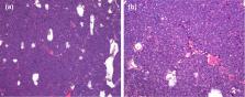
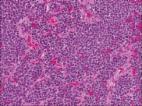
          We present a rare case of a primary intradural extramedullary Ewing’s sarcoma/peripheral primitive neuroectodermal tumor (PIEES/PNET) in the thoracolumbar spine and review the current literature. We describe the imaging manifestations, pathological features, surgical methods, and patient survival to shed light on the clinical management of this rare tumor. A 32-year-old man experienced progressive low back pain for more than 1 month. An intradural extramedullary tumor from T12 to L2 was detected on magnetic resonance imaging. He underwent a thoracolumbar laminotomy for decompression, complete excision of the intradural extramedullary tumor, and internal fixation with pedicle screws. A histopathological examination confirmed that the tumor was a PIEES/PNET via an immunohistochemical study of the surgically resected tissues. Postoperatively, the patient received chemotherapy and radiotherapy. No recurrence, metastasis, or failure of internal fixation were noted at a 17-month post-surgery radiographic examination. PIEES/PNET of the thoracolumbar spine is extremely rare. Treatment is difficult because the current literature is sparse and cases are rare. Complete resection combined with chemotherapy and radiotherapy effectively reduces recurrence and metastasis.

          Related collections

          Most cited references23

          • Record: found
          • Abstract: found
          • Article: not found

          A rare case of intradural extramedullary Ewing's sarcoma with skip metastasis in the spine.

          Spinal cord tumors are rare and fortunately affect only a minority of the population. These tumors are classified based on their anatomic location in relation to the dura mater and spinal cord as epidural, intradural extramedullary or intradural intramedullary. Intradural extramedullary Ewing's sarcoma is extremely rare. Here, we are reporting a case of intradural extramedullary Ewing's sarcoma. This is the fourth such case to be reported in English literature. A 26-year-old gentleman presented to us with low backache and progressive paraparesis from a spinal tumor. Magnetic resonance imaging showed multiple intradural, extramedullary mass lesions extending from the T11 to S2 level. Neither osteolytic nor osteosclerotic changes were seen in the vertebral bodies. Extraskeletal Ewing's sarcoma was diagnosed histopathologically. He was treated by surgery, local radiotherapy and chemotherapy. Two months after treatment, he developed a new skip lesion in the spine at T6-T7 level. The new lesion was treated with local radiotherapy and chemotherapy. Presently, the patient has completed treatment and is clinically doing fine. Intradural extramedullary Ewing's sarcoma is a rare aggressive neoplasm with high propensity for skip metastasis.
            Bookmark
            • Record: found
            • Abstract: found
            • Article: found
            Is Open Access

            Two Cases of Spinal, Extraosseous, Intradural Ewing's sarcoma/Peripheral Neuroectodermal Tumor: Radiologic, Pathologic, and Molecular Analysis

            Extraosseous Ewing's sarcoma/peripheral neuroectodermal tumors (ES/PNETs) are rare neoplasms that account for approximately 10%-15% of soft tissue sarcomas in children and 5% of soft tissue sarcomas in adults. Primary spinal, extraosseous, intradural ES/PNETs are even less common. The diagnosis of ES/PNET is extremely challenging, because the tumor can have a nonspecific radiologic appearance, and the histologic features are shared by many other “small round cell tumors.” Thus, ES/PNET should be included in the radiologic and pathologic differential diagnosis, even in older patients and in unusual tumor sites. We report two cases of spinal, extraosseous, intradural ES/PNETs in adults who presented with back pain. Magnetic resonance imaging revealed contrast enhancing, intradural lesions in the area of the conus medullaris. The tumor in Case 1 was partially intramedullary, while the tumor in Case 2 was exclusively extramedullary. In both cases, the radiologic and intraoperative surgical impression favored ependymoma. The diagnosis of ES/PNET was established in both cases by histopathologic, immunohistochemical, and molecular analysis.
              Bookmark
              • Record: found
              • Abstract: found
              • Article: not found

              Primary Intradural Ewing’s Sarcoma of The Spine: A Systematic Review of The Literature

              The incidence of extraosseous Ewing's sarcoma, a highly malignant mesenchymal mass, is rare in the spinal cord and its clinical outcomes unknown. To date, few cases of primary intradural extramedullary Ewing's sarcoma (PIEES) have been reported, with varying follow-up. Herein, we aimed to perform a comprehensive review of all cases published in the literature and update previously reported cases from our institution. Institutional medical records were searched for cases of PIEES of the spine managed at our institution between the years 2005 and 2018. We performed a systematic search of two electronic databases (Ovid Medline and Embase) from inception to August 2018 to obtain all published cases of primary intradural extraosseous Ewing's sarcoma (PIEES).We used our institutional medical records to update cases reported from our institution. We identified a total of 44 cases with pIEES reported in the existing literature of which 5 have been from our department. Of the 41 cases, the median age of diagnosis was 31 years. The most common presentation pattern was PIEES in the lumbar/sacral region (61%, n = 27), with a majority (59%, n = 25) presenting initially with pain. The most common modality of treatment reported was surgery (41/41, 100%), followed by adjuvant chemotherapy (31/44, 70%) and local radiation therapy (29/44, 66%). Overall, recurrence was reported in 17/36 (46%) cases, with median progression free survival (PFS) of 12 months (range, 1-72). There were 12/37 (32.4%) deaths reported, with median overall survival (OS) of 14 months (range, 1-72). Hence, we present the most updated review of all reported cases of PIEES. While surgical resection is the mainstay of treatment, tumor recurrence is a great concern given the adhesive nature of the lesion preventing complete resection. Adjuvant chemotherapy and radiotherapy should be carefully considered to prevent recurrence and improve survival outcome.
                Bookmark

                Author and article information

                Contributors
                Journal
                Open Med (Wars)
                Open Med (Wars)
                med
                Open Medicine
                De Gruyter
                2391-5463
                21 October 2021
                2021
                : 16
                : 1
                : 1591-1596
                Affiliations
                Department of Orthopedics, Union Hospital, Tongji Medical College, Huazhong University of Science and Technology , Wuhan 430022, China
                Department of Pathology, Union Hospital, Tongji Medical College, Huazhong University of Science and Technology , Wuhan 430022, China
                Department of Orthopedics, Union Hospital, Tongji Medical College, Huazhong University of Science and Technology , No. 1277, Jiefang Avenue, Jianghan Distinct, Wuhan 430022, China
                Author notes
                [1]

                These authors have made equal contributions to the work.

                tel: +86-27-8535-1626, fax: +86-27-8535-1626
                Article
                med-2021-0377
                10.1515/med-2021-0377
                8532567
                34722894
                572c08c6-8383-4432-8fb1-913edef81de5
                © 2021 Feifei Pu et al., published by De Gruyter

                This work is licensed under the Creative Commons Attribution 4.0 International License.

                History
                : 02 June 2021
                : 29 September 2021
                : 04 October 2021
                Page count
                Pages: 6
                Categories
                Case Report

                chemotherapy,piees/pnet,radiotherapy,surgical resection,thoracolumbar

                Comments

                Comment on this article