5
views
0
recommends
+1 Recommend
0 collections
    0
    shares
      • Record: found
      • Abstract: found
      • Article: found
      Is Open Access

      Free-breathing non-contrast flow-independent cardiovascular magnetic resonance angiography using cardiac gated, magnetization-prepared 3D Dixon method: assessment of thoracic vasculature in congenital heart disease

      research-article

      Read this article at

      Bookmark
          There is no author summary for this article yet. Authors can add summaries to their articles on ScienceOpen to make them more accessible to a non-specialist audience.

          Abstract

          Background

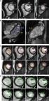
          To evaluate a non-contrast respiratory- and electrocardiogram-gated 3D cardiovascular magnetic resonance angiography (CMRA) based on magnetization-prepared Dixon method (relaxation-enhanced angiography without contrast and triggering, REACT) for the assessment of the thoracic vasculature in congenital heart disease (CHD) patients.

          Methods

          70 patients with CHD (mean 28 years, range: 10–65 years) were retrospectively identified in this single-center study. REACT-CMRA was applied with respiratory- and cardiac-gating. Image quality (IQ) of REACT-CMRA was compared to standard non-gated multi-phase first-pass-CMRA and respiratory- and electrocardiogram-gated steady-state-CMRA. IQ of different vessels of interest (ascending aorta, left pulmonary artery, left superior pulmonary vein, right coronary ostium, coronary sinus) was independently assessed by two readers on a five-point Likert scale. Measurements of vessel diameters were performed in predefined anatomic landmarks (ascending aorta, left pulmonary artery, left superior pulmonary vein). Both readers assessed artifacts and vascular abnormalities. Friedman test, chi-squared test, and Bland-Altman method were used for statistical analysis.

          Results

          Overall IQ score of REACT-CMRA was higher compared to first-pass-CMRA (3.5 ± 0.4 vs. 2.7 ± 0.4, P < 0.001) and did not differ from steady-state-CMRA (3.5 ± 0.4 vs. 3.5 ± 0.6, P = 0.99). Non-diagnostic IQ of the defined vessels of interest was observed less frequently on REACT-CMRA (1.7 %) compared to steady-state- (4.3 %, P = 0.046) or first-pass-CMRA (20.9 %, P < 0.001). Close agreements in vessel diameter measurements were observed between REACT-CMRA and steady-state-CMRA (e.g. ascending aorta, bias: 0.38 ± 1.0 mm, 95 % limits of agreement (LOA): − 1.62–2.38 mm). REACT-CMRA showed high intra- (bias: 0.04 ± 1.0 mm, 95 % LOA: − 1.9–2.0 mm) and interobserver (bias: 0.20 ± 1.1 mm, 95 % LOA: − 2.0–2.4 mm) agreements regarding vessel diameter measurements. Fat-water separation artifacts were observed in 11/70 (16 %) patients on REACT-CMRA but did not limit diagnostic utility. Six vascular abnormalities were detected on REACT-CMRA that were not seen on standard contrast-enhanced CMRA.

          Conclusions

          Non-contrast-enhanced cardiac-gated REACT-CMRA offers a high diagnostic quality for assessment of the thoracic vasculature in CHD patients.

          Related collections

          Most cited references32

          • Record: found
          • Abstract: not found
          • Article: not found

          2020 ESC Guidelines for the management of adult congenital heart disease

            Bookmark
            • Record: found
            • Abstract: found
            • Article: found
            Is Open Access

            Reference ranges (“normal values”) for cardiovascular magnetic resonance (CMR) in adults and children: 2020 update

            Cardiovascular magnetic resonance (CMR) enables assessment and quantification of morphological and functional parameters of the heart, including chamber size and function, diameters of the aorta and pulmonary arteries, flow and myocardial relaxation times. Knowledge of reference ranges (“normal values”) for quantitative CMR is crucial to interpretation of results and to distinguish normal from disease. Compared to the previous version of this review published in 2015, we present updated and expanded reference values for morphological and functional CMR parameters of the cardiovascular system based on the peer-reviewed literature and current CMR techniques. Further, databases and references for deep learning methods are included.
              Bookmark
              • Record: found
              • Abstract: found
              • Article: not found

              Gadolinium deposition in the brain: summary of evidence and recommendations.

              Emerging evidence has linked MRI signal changes in deep nuclei of the brain with repeated administration of gadolinium-based contrast agents. Gadolinium deposits have been confirmed in brain tissue, most notably in the dentate nuclei and globus pallidus. Although some linear contrast agents appear to cause greater MRI signal changes than some macrocyclic agents, deposition of gadolinium has also been observed with macrocyclic agents. However, the extent of gadolinium deposition varies between agents. Furthermore, the clinical significance of the retained gadolinium in the brain, if any, remains unknown. No data are available in human beings or animals to show adverse clinical effects due to the gadolinium deposition in the brain. On behalf of the International Society for Magnetic Resonance in Medicine, we present recommendations for the clinical and research use of gadolinium-based contrast agents. These recommendations might evolve as new evidence becomes available.
                Bookmark

                Author and article information

                Contributors
                alexander.isaak@ukbonn.de
                Journal
                J Cardiovasc Magn Reson
                J Cardiovasc Magn Reson
                Journal of Cardiovascular Magnetic Resonance
                BioMed Central (London )
                1097-6647
                1532-429X
                19 July 2021
                19 July 2021
                2021
                : 23
                : 91
                Affiliations
                [1 ]GRID grid.15090.3d, ISNI 0000 0000 8786 803X, Department of Diagnostic and Interventional Radiology, , University Hospital Bonn, ; Venusberg-Campus 1, 53127 Bonn, Germany
                [2 ]Quantitative Imaging Lab Bonn (QILaB), Bonn, Germany
                [3 ]Philips Healthcare, Hamburg, Germany
                Author information
                http://orcid.org/0000-0003-2016-6166
                Article
                788
                10.1186/s12968-021-00788-3
                8287681
                34275486
                4bb47c58-0b81-4a94-bccf-790a2f05f37f
                © The Author(s) 2021

                Open AccessThis article is licensed under a Creative Commons Attribution 4.0 International License, which permits use, sharing, adaptation, distribution and reproduction in any medium or format, as long as you give appropriate credit to the original author(s) and the source, provide a link to the Creative Commons licence, and indicate if changes were made. The images or other third party material in this article are included in the article's Creative Commons licence, unless indicated otherwise in a credit line to the material. If material is not included in the article's Creative Commons licence and your intended use is not permitted by statutory regulation or exceeds the permitted use, you will need to obtain permission directly from the copyright holder. To view a copy of this licence, visit http://creativecommons.org/licenses/by/4.0/. The Creative Commons Public Domain Dedication waiver ( http://creativecommons.org/publicdomain/zero/1.0/) applies to the data made available in this article, unless otherwise stated in a credit line to the data.

                History
                : 5 April 2021
                : 9 June 2021
                Funding
                Funded by: Universitätsklinikum Bonn (8930)
                Categories
                Research
                Custom metadata
                © The Author(s) 2021

                Cardiovascular Medicine
                congenital heart disease,thoracic vasculature,magnetic resonance angiography,non-contrast enhanced magnetic resonance angiography,react,flow-independent,free-breathing

                Comments

                Comment on this article