108
views
0
recommends
+1 Recommend
0 collections
    0
    shares
      • Record: found
      • Abstract: found
      • Article: found
      Is Open Access

      Endometrial regenerative cells: A novel stem cell population

      research-article

      Read this article at

      Bookmark
          There is no author summary for this article yet. Authors can add summaries to their articles on ScienceOpen to make them more accessible to a non-specialist audience.

          Abstract

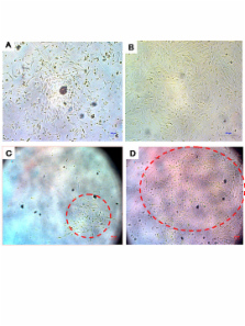
          Angiogenesis is a critical component of the proliferative endometrial phase of the menstrual cycle. Thus, we hypothesized that a stem cell-like population exist and can be isolated from menstrual blood. Mononuclear cells collected from the menstrual blood contained a subpopulation of adherent cells which could be maintained in tissue culture for >68 doublings and retained expression of the markers CD9, CD29, CD41a, CD44, CD59, CD73, CD90 and CD105, without karyotypic abnormalities. Proliferative rate of the cells was significantly higher than control umbilical cord derived mesenchymal stem cells, with doubling occurring every 19.4 hours. These cells, which we termed "Endometrial Regenerative Cells" (ERC) were capable of differentiating into 9 lineages: cardiomyocytic, respiratory epithelial, neurocytic, myocytic, endothelial, pancreatic, hepatic, adipocytic, and osteogenic. Additionally, ERC produced MMP3, MMP10, GM-CSF, angiopoietin-2 and PDGF-BB at 10–100,000 fold higher levels than two control cord blood derived mesenchymal stem cell lines. Given the ease of extraction and pluripotency of this cell population, we propose ERC as a novel alternative to current stem cells sources.

          Related collections

          Most cited references21

          • Record: found
          • Abstract: found
          • Article: not found

          Oocyte generation in adult mammalian ovaries by putative germ cells in bone marrow and peripheral blood.

          It has been suggested that germline stem cells maintain oogenesis in postnatal mouse ovaries. Here we show that adult mouse ovaries rapidly generate hundreds of oocytes, despite a small premeiotic germ cell pool. In considering the possibility of an extragonadal source of germ cells, we show expression of germline markers in bone marrow (BM). Further, BM transplantation restores oocyte production in wild-type mice sterilized by chemotherapy, as well as in ataxia telangiectasia-mutated gene-deficient mice, which are otherwise incapable of making oocytes. Donor-derived oocytes are also observed in female mice following peripheral blood transplantation. Although the fertilizability and developmental competency of the BM and peripheral blood-derived oocytes remain to be established, their morphology, enclosure within follicles, and expression of germ-cell- and oocyte-specific markers collectively support that these cells are bona fide oocytes. These results identify BM as a potential source of germ cells that could sustain oocyte production in adulthood.
            Bookmark
            • Record: found
            • Abstract: found
            • Article: not found

            Isolation and characterization of a stem cell population from adult human liver.

            Several studies suggested the presence of stem cells in the adult normal human liver; however, a population with stem cell properties has not yet been isolated. The purpose of the present study was to identify and characterize progenitor cells in normal adult human liver. By stringent conditions of liver cell cultures, we isolated and characterized a population of human liver stem cells (HLSCs). HLSCs expressed the mesenchymal stem cell markers CD29, CD73, CD44, and CD90 but not the hematopoietic stem cell markers CD34, CD45, CD117, and CD133. HLSCs were also positive for vimentin and nestin, a stem cell marker. The absence of staining for cytokeratin-19, CD117, and CD34 indicated that HLSCs were not oval stem cells. In addition, HLSCs expressed albumin, alpha-fetoprotein, and in a small percentage of cells, cytokeratin-8 and cytokeratin-18, indicating a partial commitment to hepatic cells. HLSCs differentiated in mature hepatocytes when cultured in the presence of hepatocyte growth factor and fibroblast growth factor 4, as indicated by the expression of functional cytochrome P450, albumin, and urea production. Under this condition, HLSCs downregulated alpha-fetoprotein and expressed cytokeratin-8 and cytokeratin-18. HLSCs were also able to undergo osteogenic and endothelial differentiation when cultured in the appropriated differentiation media, but they did not undergo lipogenic differentiation. Moreover, HLSCs differentiated in insulin-producing islet-like structures. In vivo, HLSCs contributed to regeneration of the liver parenchyma in severe-combined immunodeficient mice. In conclusion, we here identified a pluripotent progenitor population in adult human liver that could provide a basis for cell therapy strategies.
              Bookmark
              • Record: found
              • Abstract: found
              • Article: not found

              Embryonic stem cell immunogenicity increases upon differentiation after transplantation into ischemic myocardium.

              We investigated whether differentiation of embryonic stem cells (ESCs) in ischemic myocardium enhances their immunogenicity, thereby increasing their chance for rejection. In one series, 129/SvJ-derived mouse ESCs (ES-D3 line) were transplanted by direct myocardial injection (1 x 10(6) cells) into murine hearts of both allogeneic (BALB/c, n=20) and syngeneic (129/SvJ, n=12) recipients after left anterior artery ligation. Hearts were procured at 1, 2, 4, and 8 weeks after ESC transplantation and analyzed by immunohistochemistry to assess immune cell infiltration (CD3, CD4, CD8, B220, CD11c, Mac-1, and Gr-1) and ESC differentiation (hematoxylin and eosin). In a second series (allogeneic n=5, sham n=3), ESC transplantation was performed similarly; however after 2 weeks, left anterior descending artery-ligated and ESC-injected hearts were heterotopically transplanted into naive BALB/c recipients. After an additional 2 weeks, donor hearts were procured and analyzed by immunohistochemistry. In the first series, the size of all ESC grafts remained stable and there was no evidence of ESC differentiation 2 weeks after transplantation; however, after 4 weeks, both allogeneic and syngeneic ESC grafts showed the presence of teratoma. By 8 weeks, surviving ESCs could be detected in the syngeneic but not in the allogeneic group. Mild inflammatory cellular infiltrates were found in allogeneic recipients at 1 and 2 weeks after transplantation, progressing into vigorous infiltration at 4 and 8 weeks. The second series demonstrated similar vigorous infiltration of immune cells as early as 2 weeks after heterotopic transplantation. In vivo differentiated ESCs elicit an accelerated immune response as compared with undifferentiated ESCs. These data imply that clinical transplantation of allogeneic ESCs or ESC derivatives for treatment of cardiac failure might require immunosuppressive therapy.
                Bookmark

                Author and article information

                Journal
                J Transl Med
                Journal of Translational Medicine
                BioMed Central
                1479-5876
                2007
                15 November 2007
                : 5
                : 57
                Affiliations
                [1 ]Bio-Communications Research Institute, Wichita, USA
                [2 ]Medistem Laboratories Inc, Tempe, USA
                [3 ]Department of Surgery, University of Western Ontario, London, Canada
                [4 ]Department of Pediatrics, University of Alberta, Edmonton, Canada
                Article
                1479-5876-5-57
                10.1186/1479-5876-5-57
                2212625
                18005405
                484b9ea9-05bc-4574-bfca-23aa67d7b30d
                Copyright © 2007 Meng et al; licensee BioMed Central Ltd.

                This is an Open Access article distributed under the terms of the Creative Commons Attribution License ( http://creativecommons.org/licenses/by/2.0), which permits unrestricted use, distribution, and reproduction in any medium, provided the original work is properly cited.

                History
                : 26 September 2007
                : 15 November 2007
                Categories
                Research

                Medicine
                Medicine

                Comments

                Comment on this article