3
views
0
recommends
+1 Recommend
0 collections
    0
    shares
      • Record: found
      • Abstract: found
      • Article: found
      Is Open Access

      The effect of pre-analytical variables on downstream application and data analysis of human endometrial biopsies

      research-article

      Read this article at

      Bookmark
          There is no author summary for this article yet. Authors can add summaries to their articles on ScienceOpen to make them more accessible to a non-specialist audience.

          Abstract

          STUDY QUESTION

          What are the effects of pre-analytical variables on the downstream analysis of patient-derived endometrial biopsies?

          SUMMARY ANSWER

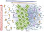
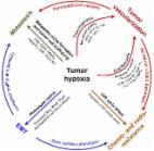
          There are distinct differences in the protein levels of the master regulator of oxygen homeostasis, hypoxia-inducible factor-1-alpha (HIF1α), and the protein and mRNA levels of three related genes, carbonic anhydrase 9 ( CA9), vascular endothelial growth factor A ( VEGFA) and progesterone receptor ( PR) in human endometrial biopsies, depending on the pre-analytical variables: disease status (cancer vs benign), timing of biopsy (pre- vs post-hysterectomy) and type of biopsy (pipelle vs full-thickness).

          WHAT IS KNOWN ALREADY

          Patient-derived biopsies are vital to endometrial research, but pre-analytical variables relating to their collection may affect downstream analysis, as is evident in other tissues.

          STUDY DESIGN, SIZE, DURATION

          A prospective observational study including patients undergoing hysterectomy for endometrial cancer (EC) or benign indications was conducted at a large tertiary gynaecological unit in the UK. Endometrial biopsies were obtained at different time points (pre- or post-hysterectomy) using either a pipelle endometrial sampler or as a full-thickness wedge biopsy.

          PARTICIPANTS/MATERIALS, SETTING, METHODS

          The changes in HIF1α, CA9, VEGFA and PR protein levels were measured by semi-quantitative analysis of immunostaining, and the expression levels of three genes ( CA9, VEGFA and PR) were investigated by quantitative real-time PCR, in endometrial biopsies from 43 patients undergoing hysterectomy for EC (n = 22) or benign gynaecological indications (n = 21).

          MAIN RESULTS AND THE ROLE OF CHANCE

          An increase in HIF1α immunostaining was observed in EC versus benign endometrium (functionalis glands) obtained pre-hysterectomy ( P < 0.001). An increase in CA9 immunostaining was observed in EC versus benign endometrial functionalis glands at both pre- and post-hysterectomy time points ( P = 0.03 and P = 0.003, respectively). Compared with benign endometrial pipelle samples, EC samples demonstrated increased mRNA expression of CA9 (pre-hysterectomy P < 0.001, post-hysterectomy P = 0.008) and VEGFA (pre-hysterectomy P = 0.004, post-hysterectomy P = 0.002). In benign uteri, HIF1α immunoscores (functionalis glands, P = 0.03 and stroma, P = 0.009), VEGFA immunoscores (functionalis glands, P = 0.03 and stroma, P = 0.01) and VEGFA mRNA levels ( P = 0.008) were increased in matched post-hysterectomy versus pre-hysterectomy samples. Similarly, in EC, an increase in VEGFA immunoscores (epithelial and stromal) and VEGFA mRNA expression was observed in the matched post-hysterectomy versus pre-hysterectomy biopsies ( P = 0.008, P = 0.004 and P = 0.018, respectively). Full-thickness benign post-hysterectomy endometrial biopsies displayed increased VEGFA ( P = 0.011) and PR ( P = 0.006) mRNA expression compared with time-matched pipelle biopsies.

          LARGE SCALE DATA

          N/A.

          LIMITATIONS, REASONS FOR CAUTION

          This descriptive study explores the effect of pre-analytical variables on the expression of four proteins and three hypoxia-related genes in a limited number of endometrial biopsies from patients with EC and benign controls. Due to the small number, it was not possible to investigate other potential variables such as menstrual cycle phase, region-specific differences within the endometrium, grade and stage of cancer, and surgical technicalities.

          WIDER IMPLICATIONS OF THE FINDINGS

          Careful consideration of the effects of these pre-analytical variables is essential when interpreting data relating to human endometrial biopsies. A standardized approach to endometrial tissue collection is essential to ensure accurate and clinically transferrable data.

          STUDY FUNDING/COMPETING INTEREST(S)

          The authors have no conflicts of interest to declare. The work included in this manuscript was funded by Wellbeing of Women project grants RG1073 and RG2137 (D.K.H.), Wellbeing of Women Entry-Level Scholarship ELS706 and Medical Research Council MR/V007238/1 (A.M./D.K.H.), Liverpool Women’s Hospital Cancer Charity (M.A.) and University of Liverpool (L.B., L.R. and E.N.).

          Related collections

          Most cited references50

          • Record: found
          • Abstract: found
          • Article: found
          Is Open Access

          The role of hypoxia in cancer progression, angiogenesis, metastasis, and resistance to therapy

          Hypoxia is a non-physiological level of oxygen tension, a phenomenon common in a majority of malignant tumors. Tumor-hypoxia leads to advanced but dysfunctional vascularization and acquisition of epithelial-to-mesenchymal transition phenotype resulting in cell mobility and metastasis. Hypoxia alters cancer cell metabolism and contributes to therapy resistance by inducing cell quiescence. Hypoxia stimulates a complex cell signaling network in cancer cells, including the HIF, PI3K, MAPK, and NFĸB pathways, which interact with each other causing positive and negative feedback loops and enhancing or diminishing hypoxic effects. This review provides background knowledge on the role of tumor hypoxia and the role of the HIF cell signaling involved in tumor blood vessel formation, metastasis, and development of the resistance to therapy. Better understanding of the role of hypoxia in cancer progression will open new windows for the discovery of new therapeutics targeting hypoxic tumor cells and hypoxic microenvironment.
            Bookmark
            • Record: found
            • Abstract: found
            • Article: not found

            HIF-1: mediator of physiological and pathophysiological responses to hypoxia.

            All organisms can sense O(2) concentration and respond to hypoxia with adaptive changes in gene expression. The large body size of mammals necessitates the development of multiple complex physiological systems to ensure adequate O(2) delivery to all cells under normal conditions. The transcriptional regulator hypoxia-inducible factor 1 (HIF-1) is an essential mediator of O(2) homeostasis. HIF-1 is required for the establishment of key physiological systems during development and their subsequent utilization in fetal and postnatal life. HIF-1 also appears to play a key role in the pathophysiology of cancer, cardiovascular disease, and chronic lung disease, which represent the major causes of mortality among industrialized societies. Genetic or pharmacological modulation of HIF-1 activity in vivo may represent a novel therapeutic approach to these disorders.
              Bookmark
              • Record: found
              • Abstract: found
              • Article: found
              Is Open Access

              The hypoxic tumour microenvironment

              Cancer progression often benefits from the selective conditions present in the tumour microenvironment, such as the presence of cancer-associated fibroblasts (CAFs), deregulated ECM deposition, expanded vascularisation and repression of the immune response. Generation of a hypoxic environment and activation of its main effector, hypoxia-inducible factor-1 (HIF-1), are common features of advanced cancers. In addition to the impact on tumour cell biology, the influence that hypoxia exerts on the surrounding cells represents a critical step in the tumorigenic process. Hypoxia indeed enables a number of events in the tumour microenvironment that lead to the expansion of aggressive clones from heterogeneous tumour cells and promote a lethal phenotype. In this article, we review the most relevant findings describing the influence of hypoxia and the contribution of HIF activation on the major components of the tumour microenvironment, and we summarise their role in cancer development and progression.
                Bookmark

                Author and article information

                Contributors
                Journal
                Hum Reprod Open
                Hum Reprod Open
                hropen
                Human Reproduction Open
                Oxford University Press
                2399-3529
                2022
                13 June 2022
                13 June 2022
                : 2022
                : 3
                : hoac026
                Affiliations
                Department of Women's and Children's Health, Institute of Life Course and Medical Sciences, University of Liverpool , Liverpool, UK
                Liverpool Women's Hospital NHS Foundation Trust, member of Liverpool Health Partners , Liverpool, UK
                Department of Women's and Children's Health, Institute of Life Course and Medical Sciences, University of Liverpool , Liverpool, UK
                Liverpool Women's Hospital NHS Foundation Trust, member of Liverpool Health Partners , Liverpool, UK
                Department of Women's and Children's Health, Institute of Life Course and Medical Sciences, University of Liverpool , Liverpool, UK
                Department of Women's and Children's Health, Institute of Life Course and Medical Sciences, University of Liverpool , Liverpool, UK
                Department of Women's and Children's Health, Institute of Life Course and Medical Sciences, University of Liverpool , Liverpool, UK
                Department of Women's and Children's Health, Institute of Life Course and Medical Sciences, University of Liverpool , Liverpool, UK
                Department of Women's and Children's Health, Institute of Life Course and Medical Sciences, University of Liverpool , Liverpool, UK
                Department of Women's and Children's Health, Institute of Life Course and Medical Sciences, University of Liverpool , Liverpool, UK
                Liverpool Women's Hospital NHS Foundation Trust, member of Liverpool Health Partners , Liverpool, UK
                Author notes
                Correspondence address. The University of Liverpool, 1st Floor University Department, Liverpool Women’s Hospital, Crown Street, Liverpool, L8 7SS, UK. Tel: +44-77-9244-3654; E-mail: a.maclean@ 123456liverpool.ac.uk
                Author information
                https://orcid.org/0000-0002-5713-6959
                https://orcid.org/0000-0003-0270-0150
                Article
                hoac026
                10.1093/hropen/hoac026
                9240853
                35775066
                70154060-5284-4e8f-bfb9-447ee0c2ef0b
                © The Author(s) 2022. Published by Oxford University Press on behalf of European Society of Human Reproduction and Embryology.

                This is an Open Access article distributed under the terms of the Creative Commons Attribution License ( https://creativecommons.org/licenses/by/4.0/), which permits unrestricted reuse, distribution, and reproduction in any medium, provided the original work is properly cited.

                History
                : 8 January 2022
                : 27 May 2022
                : 9 June 2022
                : 29 June 2022
                Page count
                Pages: 11
                Funding
                Funded by: Wellbeing of Women, DOI 10.13039/501100000325;
                Award ID: RG1073
                Award ID: RG2137
                Categories
                Original Article
                AcademicSubjects/MED00905

                pre-analytical variables,endometrial biopsy,endometrial cancer,hif1α,hypoxia

                Comments

                Comment on this article