98
views
0
recommends
+1 Recommend
0 collections
    11
    shares
      • Record: found
      • Abstract: found
      • Article: found
      Is Open Access

      In Vitro Detection of Dental Root Fractures with Cone Beam Computed Tomography (CBCT)

      research-article

      Read this article at

      Bookmark
          There is no author summary for this article yet. Authors can add summaries to their articles on ScienceOpen to make them more accessible to a non-specialist audience.

          Abstract

          Background:

          Since the diagnosis of non-displaced longitudinal fractures present difficulties for the dentist, three-dimensional evaluation is necessary.

          Objectives:

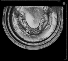
          The aim of this study is to demonstrate the accuracy of cone beam computed tomography (CBCT) in detecting dental root fractures in vitro.

          Materials and Methods:

          An in vitro model consisting of 210 recently extracted human mandibular teeth was used. Root fractures were created by mechanical force. The teeth were placed randomly in the empty dental alveoli of a dry human mandible and 15 different dental arcs were created. Images were taken with a unit Iluma ultra cone-beam CT scanner (Imtec Corporation, Germany). Three dental radiologists separately evaluated the images.

          Results:

          According to the fracture types and fracture presence, there was an overall statistically significant agreement between the key and readings. Kappa values for intra observer agreement ranged between 0.705 and 0.804 indicating that each observer gave acceptable ratings for the type and presence of fractures.

          Conclusions:

          Detailed information about root fractures may be obtained using CBCT.

          Related collections

          Most cited references38

          • Record: found
          • Abstract: found
          • Article: not found

          Clinical applications of cone-beam computed tomography in dental practice.

          Cone-beam computed tomography (CBCT) systems have been designed for imaging hard tissues of the maxillofacial region. CBCT is capable of providing sub-millimetre resolution in images of high diagnostic quality, with short scanning times (10-70 seconds) and radiation dosages reportedly up to 15 times lower than those of conventional CT scans. Increasing availability of this technology provides the dental clinician with an imaging modality capable of providing a 3-dimensional representation of the maxillofacial skeleton with minimal distortion. This article provides an overview of currently available maxillofacial CBCT systems and reviews the specific application of various CBCT display modes to clinical dental practice.
            Bookmark
            • Record: found
            • Abstract: found
            • Article: not found

            What is cone-beam CT and how does it work?

            This article on x-ray cone-beam CT (CBCT) acquisition provides an overview of the fundamental principles of operation of this technology and the influence of geometric and software parameters on image quality and patient radiation dose. Advantages of the CBCT system and a summary of the uses and limitations of the images produced are discussed. All current generations of CBCT systems provide useful diagnostic images. Future enhancements most likely will be directed toward reducing scan time; providing multimodal imaging; improving image fidelity, including soft tissue contrast; and incorporating task-specific protocols to minimize patient dose.
              Bookmark
              • Record: found
              • Abstract: found
              • Article: not found

              A new volumetric CT machine for dental imaging based on the cone-beam technique: preliminary results.

              The objective of this paper is to present a new type of volumetric CT which uses the cone-beam technique instead of traditional fan-beam technique. The machine is dedicated to the dento-maxillo-facial imaging, particularly for planning in the field of implantology. The main characteristics of the unit are presented with reference to the technical parameters as well as the software performance. Images obtained are reported as various 2D sections of a volume reconstruction. Also, measurements of the geometric accuracy and the radiation dose absorbed by the patient are obtained using specific phantoms. Absorbed dose is compared with that given off by spiral CT. Geometric accuracy, evaluated with reference to various reconstruction modalities and different spatial orientations, is 0.8-1% for width measurements and 2.2% for height measurements. Radiation dose absorbed during the scan shows different profiles in central and peripheral axes. As regards the maximum value of the central profile, dose from the new unit is approximately one sixth that of traditional spiral CT. The new system appears to be very promising in dentomaxillo-facial imaging and, due to the good ratio between performance and low cost, together with low radiation dose, very interesting in view of large-scale use of the CT technique in such diagnostic applications.
                Bookmark

                Author and article information

                Journal
                Iran J Radiol
                Iran J Radiol
                10.5812/iranjradiol
                Kowsar
                Iranian Journal of Radiology
                Kowsar
                1735-1065
                2008-2711
                30 January 2014
                January 2014
                : 11
                : 1
                : e11485
                Affiliations
                [1 ]Department of Dentomaxillofacial Radiology, Yeditepe University Faculty of Dentistry, Istanbul, Turkey
                Author notes
                [* ]Corresponding author: Erdogan Fisekcioglu, Department of Dentomaxillofacial Radiology, Yeditepe University Faculty of Dentistry, Istanbul, Turkey. Tel: +90-5325626256, Fax: +90-2163636211, E-mail: erdogan.fisekcioglu@ 123456yeditepe.edu.tr
                Article
                10.5812/iranjradiol.11485
                3955851
                460d2674-af19-4ddf-8b40-87ce00a75466
                Copyright © 2014, Tehran University of Medical Sciences and Iranian Society of Radiology; Published by Kowsar Corp.

                This is an open-access article distributed under the terms of the Creative Commons Attribution License, which permits unrestricted use, distribution, and reproduction in any medium, provided the original work is properly cited.

                History
                : 10 April 2013
                : 26 May 2013
                : 28 June 2013
                Categories
                Head & Neck Imaging

                Radiology & Imaging
                tooth fractures,in vitro,cone-beam computed tomography
                Radiology & Imaging
                tooth fractures, in vitro, cone-beam computed tomography

                Comments

                Comment on this article