5
views
0
recommends
+1 Recommend
0 collections
    0
    shares
      • Record: found
      • Abstract: found
      • Article: found
      Is Open Access

      Ischemic Heart Disease: Noninvasive Imaging Techniques and Findings

      research-article

      Read this article at

      Bookmark
          There is no author summary for this article yet. Authors can add summaries to their articles on ScienceOpen to make them more accessible to a non-specialist audience.

          Abstract

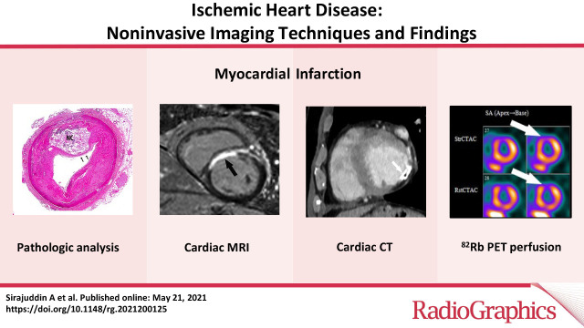
          Ischemic heart disease is a leading cause of death worldwide and comprises a large proportion of annual health care expenditure. Management of ischemic heart disease is now best guided by the physiologic significance of coronary artery stenosis. Invasive coronary angiography is the standard for diagnosing coronary artery stenosis. However, it is expensive and has risks including vascular access site complications and contrast material–induced nephropathy. Invasive coronary angiography requires fractional flow reserve (FFR) measurement to determine the physiologic significance of a coronary artery stenosis. Multiple noninvasive cardiac imaging modalities can also anatomically delineate or functionally assess for significant coronary artery stenosis, as well as detect the presence of myocardial infarction (MI). While coronary CT angiography can help assess the degree of anatomic stenosis, its inability to assess the physiologic significance of lesions limits its specificity. Physiologic significance of coronary artery stenosis can be determined by cardiac MR vasodilator or dobutamine stress imaging, CT stress perfusion imaging, FFR CT, PET myocardial perfusion imaging (MPI), SPECT MPI, and stress echocardiography. Clinically unrecognized MI, another clear indicator of physiologically significant coronary artery disease, is relatively common and is best evaluated with cardiac MRI. The authors illustrate the spectrum of imaging findings of ischemic heart disease (coronary artery disease, myocardial ischemia, and MI); highlight the advantages and disadvantages of the various noninvasive imaging methods used to assess ischemic heart disease, as illustrated by recent clinical trials; and summarize current indications and contraindications for noninvasive imaging techniques for detection of ischemic heart disease.

          Online supplemental material is available for this article.

          Published under a CC BY 4.0 license.

          Abstract

          Related collections

          Most cited references116

          • Record: found
          • Abstract: found
          • Article: not found

          Heart Disease and Stroke Statistics—2020 Update

          Circulation
            Bookmark
            • Record: found
            • Abstract: found
            • Article: not found

            Mechanisms of plaque formation and rupture.

            Atherosclerosis causes clinical disease through luminal narrowing or by precipitating thrombi that obstruct blood flow to the heart (coronary heart disease), brain (ischemic stroke), or lower extremities (peripheral vascular disease). The most common of these manifestations is coronary heart disease, including stable angina pectoris and the acute coronary syndromes. Atherosclerosis is a lipoprotein-driven disease that leads to plaque formation at specific sites of the arterial tree through intimal inflammation, necrosis, fibrosis, and calcification. After decades of indolent progression, such plaques may suddenly cause life-threatening coronary thrombosis presenting as an acute coronary syndrome. Most often, the culprit morphology is plaque rupture with exposure of highly thrombogenic, red cell-rich necrotic core material. The permissive structural requirement for this to occur is an extremely thin fibrous cap, and thus, ruptures occur mainly among lesions defined as thin-cap fibroatheromas. Also common are thrombi forming on lesions without rupture (plaque erosion), most often on pathological intimal thickening or fibroatheromas. However, the mechanisms involved in plaque erosion remain largely unknown, although coronary spasm is suspected. The calcified nodule has been suggested as a rare cause of coronary thrombosis in highly calcified and tortious arteries in older individuals. To characterize the severity and prognosis of plaques, several terms are used. Plaque burden denotes the extent of disease, whereas plaque activity is an ambiguous term, which may refer to one of several processes that characterize progression. Plaque vulnerability describes the short-term risk of precipitating symptomatic thrombosis. In this review, we discuss mechanisms of atherosclerotic plaque initiation and progression; how plaques suddenly precipitate life-threatening thrombi; and the concepts of plaque burden, activity, and vulnerability. © 2014 American Heart Association, Inc.
              Bookmark
              • Record: found
              • Abstract: found
              • Article: not found

              Fractional flow reserve versus angiography for guiding percutaneous coronary intervention.

              In patients with multivessel coronary artery disease who are undergoing percutaneous coronary intervention (PCI), coronary angiography is the standard method for guiding the placement of the stent. It is unclear whether routine measurement of fractional flow reserve (FFR; the ratio of maximal blood flow in a stenotic artery to normal maximal flow), in addition to angiography, improves outcomes. In 20 medical centers in the United States and Europe, we randomly assigned 1005 patients with multivessel coronary artery disease to undergo PCI with implantation of drug-eluting stents guided by angiography alone or guided by FFR measurements in addition to angiography. Before randomization, lesions requiring PCI were identified on the basis of their angiographic appearance. Patients assigned to angiography-guided PCI underwent stenting of all indicated lesions, whereas those assigned to FFR-guided PCI underwent stenting of indicated lesions only if the FFR was 0.80 or less. The primary end point was the rate of death, nonfatal myocardial infarction, and repeat revascularization at 1 year. The mean (+/-SD) number of indicated lesions per patient was 2.7+/-0.9 in the angiography group and 2.8+/-1.0 in the FFR group (P=0.34). The number of stents used per patient was 2.7+/-1.2 and 1.9+/-1.3, respectively (P<0.001). The 1-year event rate was 18.3% (91 patients) in the angiography group and 13.2% (67 patients) in the FFR group (P=0.02). Seventy-eight percent of the patients in the angiography group were free from angina at 1 year, as compared with 81% of patients in the FFR group (P=0.20). Routine measurement of FFR in patients with multivessel coronary artery disease who are undergoing PCI with drug-eluting stents significantly reduces the rate of the composite end point of death, nonfatal myocardial infarction, and repeat revascularization at 1 year. (ClinicalTrials.gov number, NCT00267774.) 2009 Massachusetts Medical Society
                Bookmark

                Author and article information

                Contributors
                Journal
                Radiographics
                Radiographics
                Radiographics
                Radiographics
                Radiological Society of North America
                0271-5333
                1527-1323
                21 May 2021
                July 2021
                1 July 2022
                : 41
                : 4
                : E990-E1021
                Affiliations
                [1]From the Cardiovascular and Pulmonary Branch, National Heart Lung and Blood Institute, National Institutes of Health, 10 Center Dr, Building 10, Room B1D416, Bethesda, MD 20814 (A.S., S.M.M., A.E.A.); Department of Radiology, University of California San Diego, San Diego, Calif (S.J.K.); Departments of Medicine and Radiology, Divisions of Cardiology and Cardiothoracic Imaging, University of Colorado Anschutz Medical Campus, Aurora, Colo (D.W.G.); Department of Pathology (A.P.B.) and Department of Radiology and Nuclear Medicine (C.S.W.), School of Medicine, University of Maryland, Baltimore, Md; and St David’s Healthcare and Austin Heart, Austin, Tex (F.K.).
                Author notes
                Address correspondence to A.S. (e-mail: arlene.sirajuddin@ 123456nih.gov ).
                Author information
                https://orcid.org/0000-0001-9183-5983
                https://orcid.org/0000-0002-8319-9646
                https://orcid.org/0000-0002-1532-1371
                https://orcid.org/0000-0002-4912-6320
                https://orcid.org/0000-0002-9789-7648
                https://orcid.org/0000-0002-5413-1466
                Article
                200125
                10.1148/rg.2021200125
                8262179
                34019437
                42dab303-b7bd-4c3b-98bd-b2bd68907afc
                2021 by the Radiological Society of North America, Inc.

                Published under a https://creativecommons.org/licenses/by/4.0/CC BY 4.0 license.

                History
                : 17 May 2020
                : 26 June 2020
                : 4 August 2020
                Funding
                Funded by: National Heart, Lung, and Blood Institute, National Institutes of Health
                Award ID: ZIA HL006137-10
                Categories
                Cardiac Imaging
                CA, Cardiac Radiology
                CT, Computed Tomography
                MR, Magnetic Resonance Imaging
                Custom metadata
                yes
                2024-05-21

                Comments

                Comment on this article