335
views
0
recommends
+1 Recommend
0 collections
    12
    shares
      • Record: found
      • Abstract: found
      • Article: found
      Is Open Access

      Localization of NG2 immunoreactive neuroglia cells in the rat locus coeruleus and their plasticity in response to stress

      research-article

      Read this article at

      Bookmark
          There is no author summary for this article yet. Authors can add summaries to their articles on ScienceOpen to make them more accessible to a non-specialist audience.

          Abstract

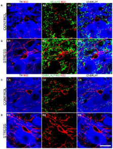
          The locus coeruleus (LC) nucleus modulates adaptive behavioral responses to stress and dysregulation of LC neuronal activity is implicated in stress-induced mental illnesses. The LC is composed primarily of noradrenergic neurons together with various glial populations. A neuroglia cell-type largely unexplored within the LC is the NG2 cell. NG2 cells serve primarily as oligodendrocyte precursor cells throughout the brain. However, some NG2 cells are in synaptic contact with neurons suggesting a role in information processing. The aim of this study was to neurochemically and anatomically characterize NG2 cells within the rat LC. Furthermore, since NG2 cells have been shown to proliferate in response to traumatic brain injury, we investigated whether such NG2 cells plasticity also occurs in response to emotive insults such as stress. Immunohistochemistry and confocal microscopy revealed that NG2 cells were enriched within the pontine region occupied by the LC. Close inspection revealed that a sub-population of NG2 cells were located within unique indentations of LC noradrenergic somata and were immunoreactive for the neuronal marker NeuN whilst NG2 cell processes formed close appositions with clusters immunoreactive for the inhibitory synaptic marker proteins gephyrin and the GABA-A receptor alpha3-subunit, on noradrenergic dendrites. In addition, LC NG2 cell processes were decorated with vesicular glutamate transporter 2 immunoreactive puncta. Finally, 10 days of repeated restraint stress significantly increased the density of NG2 cells within the LC. The study demonstrates that NG2 IR cells are integral components of the LC cellular network and they exhibit plasticity as a result of emotive challenges.

          Related collections

          Most cited references60

          • Record: found
          • Abstract: found
          • Article: not found

          NG2-expressing glial progenitor cells: an abundant and widespread population of cycling cells in the adult rat CNS.

          M. Dawson (2003)
          Glial progenitor cells of the developing CNS committed to the oligodendrocyte lineage (OPCs) express the chondroitin sulfate proteoglycan, NG2. A proportion of OPCs fail to differentiate past the stage at which they express NG2 and the lipid antigen O4 and persist in the adult CNS in a phenotypically immature form. However, the physiological function of NG2(+) cells in the adult CNS is unknown. Using antibodies against NG2 we show that NG2 is expressed by a distinct cell population in the mature CNS with the homogeneous antigenic phenotype of oligodendrocyte progenitors. The morphology of NG2(+) OPCs varies from region to region, reflecting the different structural environments, but they appear to represent a homogeneous population within any one gray or white matter region. A study of nine CNS regions showed that NG2(+) OPCs are numerous throughout the CNS and numbers in the white matter are only 1.5 times that in the gray. Whereas the ratio of OPCs to myelinating oligodendrocytes in the spinal cord gray and white matter approximates 1:4, gray matter regions of the forebrain have a 1:1 ratio, a phenomenon that will have consequences for oligodendrocyte replacement following demyelination. BrdU incorporation experiments showed that NG2(+) cells are the major dividing cell population of the adult rat CNS. Since very little apoptosis was detected and BrdU became increasingly present in oligodendrocytes after a 10-day pulse chase, with a concomitant decrease in NG2(+) BrdU incorporating cells, we suggest that the size of the oligodendrocyte population may actually increase during adult life.
            Bookmark
            • Record: found
            • Abstract: found
            • Article: not found

            Glutamatergic synapses on oligodendrocyte precursor cells in the hippocampus.

            Fast excitatory neurotransmission in the central nervous system occurs at specialized synaptic junctions between neurons, where a high concentration of glutamate directly activates receptor channels. Low-affinity AMPA (alpha-amino-3-hydroxy-5-methyl isoxazole propionic acid) and kainate glutamate receptors are also expressed by some glial cells, including oligodendrocyte precursor cells (OPCs). However, the conditions that result in activation of glutamate receptors on these non-neuronal cells are not known. Here we report that stimulation of excitatory axons in the hippocampus elicits inward currents in OPCs that are mediated by AMPA receptors. The quantal nature of these responses and their rapid kinetics indicate that they are produced by the exocytosis of vesicles filled with glutamate directly opposite these receptors. Some of these AMPA receptors are permeable to calcium ions, providing a link between axonal activity and internal calcium levels in OPCs. Electron microscopic analysis revealed that vesicle-filled axon terminals make synaptic junctions with the processes of OPCs in both the young and adult hippocampus. These results demonstrate the existence of a rapid signalling pathway from pyramidal neurons to OPCs in the mammalian hippocampus that is mediated by excitatory, glutamatergic synapses.
              Bookmark
              • Record: found
              • Abstract: found
              • Article: not found

              Convergent regulation of locus coeruleus activity as an adaptive response to stress.

              Although hypothalamic-pituitary-adrenal axis activation is generally considered to be the hallmark of the stress response, many of the same stimuli that initiate this response also activate the locus coeruleus-norepinephrine system. Given its functional attributes, the parallel engagement of the locus coeruleus-norepinephrine system with the hypothalamic-pituitary-adrenal axis serves to coordinate endocrine and cognitive limbs of the stress response. The elucidation of stress-related afferents to the locus coeruleus and the electrophysiological characterization of these inputs are revealing how the activity of this system is fine-tuned by stressors to facilitate adaptive cognitive responses. Emerging from these studies, is a picture of complex interactions between the stress-related neuropeptide, corticotropin-releasing factor (CRF), endogenous opioids and the excitatory amino acid neurotransmitter, glutamate. The net effect of these interactions is to adjust the activity and reactivity of the locus coeruleus-norepinephrine system such that state of arousal and processing of sensory stimuli are modified to facilitate adaptive behavioral responses to stressors. This review begins with an introduction to the basic anatomical and physiological characteristics of locus coeruleus neurons. The concept that locus coeruleus neurons operate through two activity modes, i.e., tonic vs. phasic, that determine distinct behavioral strategies is emphasized in light of its relevance to stress. Anatomical and physiological evidence are then presented suggesting that interactions between stress-related neurotransmitters that converge on locus coeruleus neurons regulate shifts between these modes of discharge in response to the challenge of a stressor. This review focuses specifically on the locus coeruleus because it is the major source of norepinephrine to the forebrain and has been implicated in behavioral and cognitive aspects of stress responses.
                Bookmark

                Author and article information

                Contributors
                Journal
                Front Neuroanat
                Front Neuroanat
                Front. Neuroanat.
                Frontiers in Neuroanatomy
                Frontiers Media S.A.
                1662-5129
                14 May 2014
                2014
                : 8
                : 31
                Affiliations
                [1] 1Institute for Biomedical and Biomolecular Sciences, School of Pharmacy and Biomedical Sciences, University of Portsmouth Portsmouth, UK
                [2] 2Department of Cell Biology, University Medical Centre Groningen, University of Groningen Groningen, Netherlands
                [3] 3Electron Microscopy and Histology, Department of Laboratory Medicine, Children's and Women's Health, Norwegian University of Science and Technology Trondheim, Norway
                [4] 4Pharma Research and Early Development, DTA Neuroscience, F. Hoffmann-La Roche Ltd Basel, Switzerland
                Author notes

                Edited by: Paul J. May, University of Mississippi Medical Center, USA

                Reviewed by: Yugo Fukazawa, Nagoya University Graduate School of Medicine, Japan; Rick C. S. Lin, University of Mississippi Medical Center, USA

                *Correspondence: Jerome D. Swinny, Institute for Biomedical and Biomolecular Sciences, School of Pharmacy and Biomedical Sciences, University of Portsmouth, St Michael's Building, White Swan Road, Portsmouth PO1 2DT, UK e-mail: jerome.swinny@ 123456port.ac.uk

                Mohsen Seifi and Nicole L. Corteen have contributed equally to this work.

                This article was submitted to the journal Frontiers in Neuroanatomy.

                Article
                10.3389/fnana.2014.00031
                4030166
                39f6f745-09b9-4d30-89e3-b74e6a03863a
                Copyright © 2014 Seifi, Corteen, van der Want, Metzger and Swinny.

                This is an open-access article distributed under the terms of the Creative Commons Attribution License (CC BY). The use, distribution or reproduction in other forums is permitted, provided the original author(s) or licensor are credited and that the original publication in this journal is cited, in accordance with accepted academic practice. No use, distribution or reproduction is permitted which does not comply with these terms.

                History
                : 24 February 2014
                : 21 April 2014
                Page count
                Figures: 8, Tables: 1, Equations: 0, References: 75, Pages: 14, Words: 0
                Categories
                Neuroscience
                Original Research Article

                Neurosciences
                opc,noradrenaline,brainstem,glia,gabaa receptor
                Neurosciences
                opc, noradrenaline, brainstem, glia, gabaa receptor

                Comments

                Comment on this article