5
views
0
recommends
+1 Recommend
0 collections
    0
    shares
      • Record: found
      • Abstract: found
      • Article: found
      Is Open Access

      Dynamic Modulation of Mouse Locus Coeruleus Neurons by Vasopressin 1a and 1b Receptors

      research-article

      Read this article at

      Bookmark
          There is no author summary for this article yet. Authors can add summaries to their articles on ScienceOpen to make them more accessible to a non-specialist audience.

          Abstract

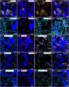
          The locus coeruleus (LC) is a brainstem nucleus distinguished by its supply of noradrenaline throughout the central nervous system. Apart from modulating a range of brain functions, such as arousal, cognition and the stress response, LC neuronal excitability also corresponds to the activity of various peripheral systems, such as pelvic viscera and the cardiovascular system. Neurochemically diverse inputs set the tone for LC neuronal activity, which in turn modulates these adaptive physiological and behavioral responses essential for survival. One such LC afferent system which is poorly understood contains the neurohormone arginine-vasopressin (AVP). Here we provide the first demonstration of the molecular and functional characteristics of the LC-AVP system, by characterizing its receptor-specific modulation of identified LC neurons and plasticity in response to stress. High resolution confocal microscopy revealed that immunoreactivity for the AVP receptor 1b (V1b) was located on plasma membranes of noradrenergic and non-noradrenergic LC neurons. In contrast, immunoreactivity for the V1a receptor was exclusively located on LC noradrenergic neurons. No specific signal, either at the mRNA or protein level, was detected for the V2 receptor in the LC. Clusters immunoreactive for V1a-b were located in proximity to profiles immunoreactive for GABAergic and glutamatergic synaptic marker proteins. AVP immunopositive varicosities were also located adjacent to labeling for such synaptic markers. Whole-cell patch clamp electrophysiology revealed that the pharmacological activation of V1b receptors significantly increased the spontaneous activity of 45% (9/20) of recorded noradrenergic neurons, with the remaining 55% (11/20) of cells exhibiting a significant decrease in their basal firing patterns. Blockade of V1a and V1b receptors on their own significantly altered LC neuronal excitability in a similar heterogeneous manner, demonstrating that endogenous AVP sets the basal LC neuronal firing rates. Finally, exposing animals to acute stress increased V1b, but not V1a receptor expression, whilst decreasing AVP immunoreactivity. This study reveals the AVP-V1a-b system as a considerable component of the LC molecular architecture and regulator of LC activity. Since AVP primarily functions as a regulator of homeostasis, the data suggest a novel pathway by modulating the functioning of a brain region that is integral to mediating adaptive responses.

          Related collections

          Most cited references64

          • Record: found
          • Abstract: not found
          • Article: not found

          Characterization of a 41-residue ovine hypothalamic peptide that stimulates secretion of corticotropin and beta-endorphin

            Bookmark
            • Record: found
            • Abstract: found
            • Article: not found

            CRH Engagement of the Locus Coeruleus Noradrenergic System Mediates Stress-Induced Anxiety.

            The locus coeruleus noradrenergic (LC-NE) system is one of the first systems engaged following a stressful event. While numerous groups have demonstrated that LC-NE neurons are activated by many different stressors, the underlying neural circuitry and the role of this activity in generating stress-induced anxiety has not been elucidated. Using a combination of in vivo chemogenetics, optogenetics, and retrograde tracing, we determine that increased tonic activity of the LC-NE system is necessary and sufficient for stress-induced anxiety and aversion. Selective inhibition of LC-NE neurons during stress prevents subsequent anxiety-like behavior. Exogenously increasing tonic, but not phasic, activity of LC-NE neurons is alone sufficient for anxiety-like and aversive behavior. Furthermore, endogenous corticotropin-releasing hormone(+) (CRH(+)) LC inputs from the amygdala increase tonic LC activity, inducing anxiety-like behaviors. These studies position the LC-NE system as a critical mediator of acute stress-induced anxiety and offer a potential intervention for preventing stress-related affective disorders.
              Bookmark
              • Record: found
              • Abstract: found
              • Article: not found

              Convergent regulation of locus coeruleus activity as an adaptive response to stress.

              Although hypothalamic-pituitary-adrenal axis activation is generally considered to be the hallmark of the stress response, many of the same stimuli that initiate this response also activate the locus coeruleus-norepinephrine system. Given its functional attributes, the parallel engagement of the locus coeruleus-norepinephrine system with the hypothalamic-pituitary-adrenal axis serves to coordinate endocrine and cognitive limbs of the stress response. The elucidation of stress-related afferents to the locus coeruleus and the electrophysiological characterization of these inputs are revealing how the activity of this system is fine-tuned by stressors to facilitate adaptive cognitive responses. Emerging from these studies, is a picture of complex interactions between the stress-related neuropeptide, corticotropin-releasing factor (CRF), endogenous opioids and the excitatory amino acid neurotransmitter, glutamate. The net effect of these interactions is to adjust the activity and reactivity of the locus coeruleus-norepinephrine system such that state of arousal and processing of sensory stimuli are modified to facilitate adaptive behavioral responses to stressors. This review begins with an introduction to the basic anatomical and physiological characteristics of locus coeruleus neurons. The concept that locus coeruleus neurons operate through two activity modes, i.e., tonic vs. phasic, that determine distinct behavioral strategies is emphasized in light of its relevance to stress. Anatomical and physiological evidence are then presented suggesting that interactions between stress-related neurotransmitters that converge on locus coeruleus neurons regulate shifts between these modes of discharge in response to the challenge of a stressor. This review focuses specifically on the locus coeruleus because it is the major source of norepinephrine to the forebrain and has been implicated in behavioral and cognitive aspects of stress responses.
                Bookmark

                Author and article information

                Contributors
                Journal
                Front Neurosci
                Front Neurosci
                Front. Neurosci.
                Frontiers in Neuroscience
                Frontiers Media S.A.
                1662-4548
                1662-453X
                10 December 2018
                2018
                : 12
                : 919
                Affiliations
                [1] 1Departamento de Fisiología, Facultad de Medicina, Universidad Nacional Autónoma de México , Mexico City, Mexico
                [2] 2Institute for Biomedical and Biomolecular Sciences, School of Pharmacy and Biomedical Sciences, University of Portsmouth , Portsmouth, United Kingdom
                [3] 3Department of Anatomy, Charité - Universitätsmedizin Berlin , Berlin, Germany
                [4] 4I.M. Sechenov First Moscow State Medical University of the Ministry of Healthcare of the Russian Federation (Sechenovskiy University) , Moscow, Russia
                [5] 5Division of Molecular Pharmacology, Department of Pharmacology, Jichi Medical University , Shimotsuke, Japan
                Author notes

                Edited by: Vincent Geenen, University of Liège, Belgium

                Reviewed by: David Murphy, University of Bristol, United Kingdom; Antoine Adamantidis, University of Bern, Switzerland

                *Correspondence: Limei Zhang, limei.zhangdebarrio@ 123456gmail.com Jerome D. Swinny, jerome.swinny@ 123456port.ac.uk

                These authors have contributed equally to this work

                Present address: Louise Kelly, Faculty of Medicine, University of Southampton, Southampton, United Kingdom

                This article was submitted to Neuroendocrine Science, a section of the journal Frontiers in Neuroscience

                Article
                10.3389/fnins.2018.00919
                6295453
                30618551
                b72ae5a2-01bb-4d78-9d99-33831b93fec9
                Copyright © 2018 Campos-Lira, Kelly, Seifi, Jackson, Giesecke, Mutig, Koshimizu, Hernandez, Zhang and Swinny.

                This is an open-access article distributed under the terms of the Creative Commons Attribution License (CC BY). The use, distribution or reproduction in other forums is permitted, provided the original author(s) and the copyright owner(s) are credited and that the original publication in this journal is cited, in accordance with accepted academic practice. No use, distribution or reproduction is permitted which does not comply with these terms.

                History
                : 28 August 2018
                : 22 November 2018
                Page count
                Figures: 9, Tables: 0, Equations: 0, References: 73, Pages: 19, Words: 0
                Categories
                Neuroscience
                Original Research

                Neurosciences
                v1b,desmopressin,patch clamp,tasp 0390325,restraint stress
                Neurosciences
                v1b, desmopressin, patch clamp, tasp 0390325, restraint stress

                Comments

                Comment on this article