0
views
0
recommends
+1 Recommend
0 collections
    0
    shares
      • Record: found
      • Abstract: found
      • Article: found
      Is Open Access

      M1 macrophage-derived exosomes inhibit cardiomyocyte proliferation through delivering miR-155

      research-article

      Read this article at

      Bookmark
          There is no author summary for this article yet. Authors can add summaries to their articles on ScienceOpen to make them more accessible to a non-specialist audience.

          Abstract

          Background

          M1 macrophages are closely associated with cardiac injury after myocardial infarction (MI). Increasing evidence shows that exosomes play a key role in pathophysiological regulation after MI, but the role of M1 macrophage-derived exosomes (M1-Exos) in myocardial regeneration remains unclear. In this study, we explored the impact of M1 macrophage-derived exosomes on cardiomyocytes regeneration in vitro and in vivo.

          Methods

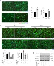
          M0 macrophages were induced to differentiate into M1 macrophages with GM-CSF (50 ng/mL) and IFN-γ (20 ng/mL). Then M1-Exos were isolated and co-incubated with cardiomyocytes. Cardiomyocyte proliferation was detected by pH3 or ki67 staining. Quantitative real-time PCR (qPCR) was used to test the level of miR-155 in macrophages, macrophage-derived exosomes and exosome-treated cardiomyocytes. MI model was constructed and LV-miR-155 was injected around the infarct area, the proliferation of cardiomyocytes was counted by pH3 or ki67 staining. The downstream gene and pathway of miR-155 were predicted and verified by dual-luciferase reporter gene assay, qPCR and immunoblotting analysis. IL-6 (50 ng/mL) was added to cardiomyocytes transfected with miR-155 mimics, and the proliferation of cardiomyocytes was calculated by immunofluorescence. The protein expressions of IL-6R, p-JAK2 and p-STAT3 were detected by Western blot.

          Results

          The results showed that M1-Exos suppressed cardiomyocytes proliferation. Meanwhile, miR-155 was highly expressed in M1-Exos and transferred to cardiomyocytes. miR-155 inhibited the proliferation of cardiomyocytes and antagonized the pro-proliferation effect of interleukin 6 (IL-6). Furthermore, miR-155 targeted gene IL-6 receptor (IL-6R) and inhibited the Janus kinase 2(JAK)/Signal transducer and activator of transcription (STAT3) signaling pathway.

          Conclusion

          M1-Exos inhibited cardiomyocyte proliferation by delivering miR-155 and inhibiting the IL-6R/JAK/STAT3 signaling pathway. This study provided new insight and potential treatment strategy for the regulation of myocardial regeneration and cardiac repair by macrophages.

          Supplementary Information

          The online version contains supplementary material available at 10.1186/s12872-024-03893-0.

          Related collections

          Most cited references24

          • Record: found
          • Abstract: not found
          • Article: not found

          Fourth Universal Definition of Myocardial Infarction (2018)

            Bookmark
            • Record: found
            • Abstract: found
            • Article: not found

            Evidence for cardiomyocyte renewal in humans.

            It has been difficult to establish whether we are limited to the heart muscle cells we are born with or if cardiomyocytes are generated also later in life. We have taken advantage of the integration of carbon-14, generated by nuclear bomb tests during the Cold War, into DNA to establish the age of cardiomyocytes in humans. We report that cardiomyocytes renew, with a gradual decrease from 1% turning over annually at the age of 25 to 0.45% at the age of 75. Fewer than 50% of cardiomyocytes are exchanged during a normal life span. The capacity to generate cardiomyocytes in the adult human heart suggests that it may be rational to work toward the development of therapeutic strategies aimed at stimulating this process in cardiac pathologies.
              Bookmark
              • Record: found
              • Abstract: found
              • Article: not found

              Revisiting Cardiac Cellular Composition.

              Accurate knowledge of the cellular composition of the heart is essential to fully understand the changes that occur during pathogenesis and to devise strategies for tissue engineering and regeneration.
                Bookmark

                Author and article information

                Contributors
                shaojunliu@gzhmu.edu.cn
                liushiming@gzhmu.edu.cn
                Journal
                BMC Cardiovasc Disord
                BMC Cardiovasc Disord
                BMC Cardiovascular Disorders
                BioMed Central (London )
                1471-2261
                16 July 2024
                16 July 2024
                2024
                : 24
                : 365
                Affiliations
                Department of Cardiology, Guangdong Key Laboratory of Vascular Diseases, The Second Affiliated Hospital, Guangzhou Institute of Cardiovascular Disease, Guangzhou Medical University, ( https://ror.org/00zat6v61) Guangzhou, 510260 People’s Republic of China
                Article
                3893
                10.1186/s12872-024-03893-0
                11251235
                39014329
                312caf09-3fab-4aa5-8e82-df831864269e
                © The Author(s) 2024

                Open Access This article is licensed under a Creative Commons Attribution 4.0 International License, which permits use, sharing, adaptation, distribution and reproduction in any medium or format, as long as you give appropriate credit to the original author(s) and the source, provide a link to the Creative Commons licence, and indicate if changes were made. The images or other third party material in this article are included in the article’s Creative Commons licence, unless indicated otherwise in a credit line to the material. If material is not included in the article’s Creative Commons licence and your intended use is not permitted by statutory regulation or exceeds the permitted use, you will need to obtain permission directly from the copyright holder. To view a copy of this licence, visit http://creativecommons.org/licenses/by/4.0/. The Creative Commons Public Domain Dedication waiver ( http://creativecommons.org/publicdomain/zero/1.0/) applies to the data made available in this article, unless otherwise stated in a credit line to the data.

                History
                : 17 November 2023
                : 16 April 2024
                Funding
                Funded by: Guangdong Basic and Applied Basic Research Foundation
                Award ID: 2022A1515012474, 2021A1515012546, 2021A1515011364, 2019A1515011214
                Award ID: 2022A1515012474, 2021A1515012546, 2021A1515011364, 2019A1515011214
                Funded by: Innovation Team of General Universities in Guangdong Province
                Award ID: 2023KCXTD025
                Award ID: 2023KCXTD025
                Funded by: Guangzhou Science and Technology Plan
                Award ID: 202102010101, 202201020220
                Award ID: 202102010101, 202201020220
                Funded by: Student Innovation Program of Guangzhou Medical University
                Award ID: to Shao.L.
                Award ID: to Shao.L.
                Categories
                Research
                Custom metadata
                © BioMed Central Ltd., part of Springer Nature 2024

                Cardiovascular Medicine
                m1 macrophage,exosomes,mir-155,myocardial infarction,cardiomyocyte proliferation

                Comments

                Comment on this article