35
views
0
recommends
+1 Recommend
0 collections
    0
    shares
      • Record: found
      • Abstract: found
      • Article: found
      Is Open Access

      Quantitative Multicolor Super-Resolution Microscopy Reveals Tetherin HIV-1 Interaction

      research-article

      Read this article at

      Bookmark
          There is no author summary for this article yet. Authors can add summaries to their articles on ScienceOpen to make them more accessible to a non-specialist audience.

          Abstract

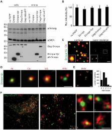
          Virus assembly and interaction with host-cell proteins occur at length scales below the diffraction limit of visible light. Novel super-resolution microscopy techniques achieve nanometer resolution of fluorescently labeled molecules. The cellular restriction factor tetherin (also known as CD317, BST-2 or HM1.24) inhibits the release of human immunodeficiency virus 1 (HIV-1) through direct incorporation into viral membranes and is counteracted by the HIV-1 protein Vpu. For super-resolution analysis of HIV-1 and tetherin interactions, we established fluorescence labeling of HIV-1 proteins and tetherin that preserved HIV-1 particle formation and Vpu-dependent restriction, respectively. Multicolor super-resolution microscopy revealed important structural features of individual HIV-1 virions, virus assembly sites and their interaction with tetherin at the plasma membrane. Tetherin localization to micro-domains was dependent on both tetherin membrane anchors. Tetherin clusters containing on average 4 to 7 tetherin dimers were visualized at HIV-1 assembly sites. Combined biochemical and super-resolution analysis revealed that extended tetherin dimers incorporate both N-termini into assembling virus particles and restrict HIV-1 release. Neither tetherin domains nor HIV-1 assembly sites showed enrichment of the raft marker GM1. Together, our super-resolution microscopy analysis of HIV-1 interactions with tetherin provides new insights into the mechanism of tetherin-mediated HIV-1 restriction and paves the way for future studies of virus-host interactions.

          Author Summary

          Human immunodeficiency virus 1 (HIV-1) assembles and interacts with cellular proteins at the plasma membrane of infected cells. Here, we analyzed individual HIV-1 virions, viral assembly sites and the mechanism of tetherin restriction by multicolor super-resolution microscopy using fully functional fluorescently labeled tetherin and viral proteins. Viral proteins within virions were visualized with nanometer resolution yielding new insight into the structure of the HIV-1. Our super-resolution analysis was extended to tetherin, a cellular restriction factor that inhibits the release of several enveloped viruses. Tetherin was localized in clusters of 70–90 nm at the plasma membrane that contain 5–11 dimers. In contrast tetherin clusters found at HIV-1 assembly sites contained on average 4–7 tetherin dimers. Clustering of tetherin was dependent on both tetherin membrane anchors. The transmembrane domain of tetherin associated with budding virions independently of GM1 lipid raft domains. Our data indicated that extended dimers tether HIV-1 virions directly to the cell. Overall, we provide for the first time super-resolution analysis of authentic virions, virus budding sites and HIV-1 interactions with the anti-viral factor tetherin. Our data offer novel insights into the mechanisms of tetherin restriction.

          Related collections

          Most cited references49

          • Record: found
          • Abstract: found
          • Article: not found

          Tetherin inhibits retrovirus release and is antagonized by HIV-1 Vpu.

          Human cells possess an antiviral activity that inhibits the release of retrovirus particles, and other enveloped virus particles, and is antagonized by the HIV-1 accessory protein, Vpu. This antiviral activity can be constitutively expressed or induced by interferon-alpha, and it consists of protein-based tethers, which we term 'tetherins', that cause retention of fully formed virions on infected cell surfaces. Using deductive constraints and gene expression analyses, we identify CD317 (also called BST2 or HM1.24), a membrane protein of previously unknown function, as a tetherin. Specifically, CD317 expression correlated with, and induced, a requirement for Vpu during HIV-1 and murine leukaemia virus particle release. Furthermore, in cells where HIV-1 virion release requires Vpu expression, depletion of CD317 abolished this requirement. CD317 caused retention of virions on cell surfaces and, after endocytosis, in CD317-positive compartments. Vpu co-localized with CD317 and inhibited these effects. Inhibition of Vpu function and consequent mobilization of tetherin's antiviral activity is a potential therapeutic strategy in HIV/AIDS.
            Bookmark
            • Record: found
            • Abstract: found
            • Article: not found

            In vivo gene delivery and stable transduction of nondividing cells by a lentiviral vector.

            A retroviral vector system based on the human immunodeficiency virus (HIV) was developed that, in contrast to a murine leukemia virus-based counterpart, transduced heterologous sequences into HeLa cells and rat fibroblasts blocked in the cell cycle, as well as into human primary macrophages. Additionally, the HIV vector could mediate stable in vivo gene transfer into terminally differentiated neurons. The ability of HIV-based viral vectors to deliver genes in vivo into nondividing cells could increase the applicability of retroviral vectors in human gene therapy.
              Bookmark
              • Record: found
              • Abstract: found
              • Article: not found

              The interferon-induced protein BST-2 restricts HIV-1 release and is downregulated from the cell surface by the viral Vpu protein.

              The HIV-1 accessory protein Vpu counteracts a host factor that restricts virion release from infected cells. Here we show that the interferon-induced cellular protein BST-2/HM1.24/CD317 is such a factor. BST-2 is downregulated from the cell surface by Vpu, and BST-2 is specifically expressed in cells that support the vpu phenotype. Exogenous expression of BST-2 inhibits HIV-1 virion release, while suppression of BST-2 relieves the requirement for Vpu. Downregulation of BST-2 requires both the transmembrane/ion channel domain and conserved serines in the cytoplasmic domain of Vpu. Endogenous BST-2 colocalizes with the HIV-1 structural protein Gag in endosomes and at the plasma membrane, suggesting that BST-2 traps virions within and on infected cells. The unusual structure of BST-2, which includes a transmembrane domain and a lumenal GPI anchor, may allow it to retain nascent enveloped virions on cellular membranes, providing a mechanism of viral restriction counteracted by a specific viral accessory protein.
                Bookmark

                Author and article information

                Contributors
                Role: Editor
                Journal
                PLoS Pathog
                plos
                plospath
                PLoS Pathogens
                Public Library of Science (San Francisco, USA )
                1553-7366
                1553-7374
                December 2011
                December 2011
                15 December 2011
                : 7
                : 12
                : e1002456
                Affiliations
                [1 ]Departments of Microbiology and Molecular Medicine, Dermatology and Venereology, University Hospital and Medical School of Geneva, Geneva, Switzerland
                [2 ]Laboratory for Photochemistry and Spectroscopy, Department of Chemistry, Katholieke Universiteit Leuven, Heverlee, Belgium
                [3 ]Department of Dermatology and Wound Healing, Cardiff University School of Medicine and University Hospital of Wales, Cardiff, Wales, United Kingdom
                Universitätsklinikum Heidelberg, Germany
                Author notes

                Conceived and designed the experiments: ML JH VP. Performed the experiments: ML SR BM FB HU. Analyzed the data: ML SR BM FB HU. Contributed reagents/materials/analysis tools: SR BM HU. Wrote the paper: ML VP. Build set-up: SR HU ML.

                Article
                PPATHOGENS-D-11-01015
                10.1371/journal.ppat.1002456
                3240612
                22194693
                2d3cd6de-6cd3-4b04-b54a-4b23e60a0c78
                Lehmann et al. This is an open-access article distributed under the terms of the Creative Commons Attribution License, which permits unrestricted use, distribution, and reproduction in any medium, provided the original author and source are credited.
                History
                : 17 May 2011
                : 9 November 2011
                Page count
                Pages: 15
                Categories
                Research Article
                Biology
                Microbiology
                Virology
                Viral Replication
                Viral Packaging
                Viral Structure
                Viral Core
                Viral Envelope
                Immunodeficiency Viruses
                Host-Pathogen Interaction
                Molecular Cell Biology
                Membranes and Sorting

                Infectious disease & Microbiology
                Infectious disease & Microbiology

                Comments

                Comment on this article