35
views
0
recommends
+1 Recommend
0 collections
    0
    shares
      • Record: found
      • Abstract: found
      • Article: found
      Is Open Access

      Correlation of Choroidal Thickness and Volume Measurements with Axial Length and Age Using Swept Source Optical Coherence Tomography and Optical Low-Coherence Reflectometry

      research-article

      Read this article at

      Bookmark
          There is no author summary for this article yet. Authors can add summaries to their articles on ScienceOpen to make them more accessible to a non-specialist audience.

          Abstract

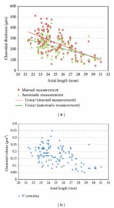
          Purpose. To report choroidal thickness and volume in healthy eyes using swept source optical coherence tomography (SS-OCT). Methods. A prospective observational study of 122 patients examined with swept source OCT (DRI-OCT, Topcon, Japan). In each eye, we performed 256 horizontal scans, 12 mm in length and centered on the fovea. We calculated choroidal thickness manually with a built-in caliper and automatically using DRI-OCT mapping software. Choroidal volume was also automatically calculated. We measured axial length with optical low-coherence reflectometry (Lenstar LS 900, Haag-Streit, Switzerland). Results. The choroid has focally increased thickness under the fovea. Choroid was thinnest in the outer nasal quadrant. In stepwise regression analysis, age was estimated as the most significant factor correlating with decreased choroidal thickness ( F = 23.146, P < 0.001) followed by axial length ( F = 4.902, P = 0.03). Refractive error was not statistically significant ( F = 1.16, P = 0.28). Conclusions. SS-OCT is the first commercially available system that can automatically create choroidal thickness and volume maps. Choroidal thickness is increased at the fovea and is thinnest nasally. Age and axial length are critical for the estimation of choroidal thickness and volume. Choroidal measurements derived from SS-OCT images have potential value for objectively documenting disease-related choroidal thickness abnormalities and monitoring progressive changes over time.

          Related collections

          Most cited references17

          • Record: found
          • Abstract: found
          • Article: not found

          Enhanced depth imaging optical coherence tomography of the choroid in central serous chorioretinopathy.

          The purpose of the study was to evaluate the choroidal thickness in patients with central serous chorioretinopathy, a disease attributed to increased choroidal vascular hyperpermeability. Patients with central serous chorioretinopathy underwent enhanced depth imaging spectral-domain optical coherence tomography, which was obtained by positioning a spectral-domain optical coherence tomography device close enough to the eye to acquire an inverted image. Seven sections, each comprising 100 averaged scans, were obtained within a 5 degrees x 30 degrees rectangle to encompass the macula. The subfoveal choroidal thickness was measured from the outer border of the retinal pigment epithelium to the inner scleral border. The mean age of subjects undergoing enhanced depth imaging spectral-domain optical coherence tomography was 59.3 years (standard deviation, 15.8 years). Seventeen of 19 patients (89.5%) were men, and 12 (63.2%) patients had bilateral clinical disease. The choroidal thickness measured in 28 eligible eyes of the 19 patients was 505 microm (standard deviation, 124 microm), which was significantly greater than the choroidal thickness in normal eyes (P < or = 0.001). Enhanced depth imaging spectral-domain optical coherence tomography demonstrated a very thick choroid in patients with central serous chorioretinopathy. This finding provides additional evidence that central serous chorioretinopathy may be caused by increased hydrostatic pressure in the choroid.
            Bookmark
            • Record: found
            • Abstract: found
            • Article: not found

            Enhanced depth imaging spectral-domain optical coherence tomography.

            To describe a method to obtain images of the choroid using conventional spectral-domain (SD) optical coherence tomography (OCT) and to evaluate choroidal thickness measurements using these images. Observational case series. The images were obtained by positioning the SD OCT device close enough to the eye to obtain an inverted representation of the fundus in healthy volunteers who did not have pupillary dilation. Seven sections, each comprised of 100 averaged scans, were obtained within a 5- x 15-degree rectangle centered on the fovea. The choroidal thickness under the fovea in each image was measured by independent observers. The choroidal thickness could be evaluated in every subject's choroidal image. The mean choroidal thickness under the fovea was 318 microm in the right eye and 335 microm in the left eye. The choroidal thickness showed a high correlation in both eyes (r = 0.82; P < .001). The correlation between the measurements performed by the independent observers was highly significant (right eye, r = 0.93; left eye, r = 0.97; P < .001 for both). This method provides detailed, measurable images from the choroid, a structure that heretofore has been difficult to image in clinical practice.
              Bookmark
              • Record: found
              • Abstract: found
              • Article: not found

              Morphometric analysis of Bruch's membrane, the choriocapillaris, and the choroid in aging.

              To quantify changes in choriocapillary density and in thickness of Bruch's membrane, the choriocapillaris, and the choroid in 95 unpaired, histologically normal human maculae aged 6 to 100 years and in 25 maculae with advanced age-related macular degeneration. Light microscopic, computer-aided, morphometric quantitative analysis. In ten decades, Bruch's membrane thickness increased by 135%, from 2.0 to 4.7 microns; the choriocapillary density decreased by 45%; the diameter of the choriocapillaris decreased by 34%, from 9.8 to 6.5 microns; and the choroidal thickness decreased by 57%, from 193.5 to 84 microns in normal maculae. In maculae with basal laminar deposit, geographic atrophy, or disciform scarring, the density of the choriocapillaris was 63%, 54%, and 43% of normal and the choriocapillary diameter was 81%, 73%, and 75% of normal, respectively. Choroidal thickness remained unchanged. Thickness of Bruch's membrane was only related to age (rs = 0.63) and not to age-related atrophy of the choriocapillaris. Age was also the strongest factor related to choriocapillary density (rs = -0.58). In advanced stages of age-related macular degeneration, the decrease in choriocapillary density and diameter was significantly larger than in normal maculae, but the thickness of the choroid and Bruch's membrane was the same. The latter was significantly thinner (81% of normal) in disciform scarring.
                Bookmark

                Author and article information

                Journal
                Biomed Res Int
                Biomed Res Int
                BMRI
                BioMed Research International
                Hindawi Publishing Corporation
                2314-6133
                2314-6141
                2014
                12 June 2014
                : 2014
                : 639160
                Affiliations
                Ophthalmic Clinic Jasne Blonia, ul. Rojna 90, 91-162 Lodz, Poland
                Author notes
                *Zofia Michalewska: zosia_n@ 123456yahoo.com

                Academic Editor: Ron Adelman

                Author information
                http://orcid.org/0000-0001-8376-9218
                Article
                10.1155/2014/639160
                4075071
                25013793
                2b7dbde2-398b-471f-8a76-f548520037c3
                Copyright © 2014 Janusz Michalewski et al.

                This is an open access article distributed under the Creative Commons Attribution License, which permits unrestricted use, distribution, and reproduction in any medium, provided the original work is properly cited.

                History
                : 26 February 2014
                : 20 May 2014
                Categories
                Research Article

                Comments

                Comment on this article