44
views
0
recommends
+1 Recommend
0 collections
    0
    shares
      • Record: found
      • Abstract: found
      • Article: found
      Is Open Access

      Pachychoroid Diseases of the Macula

      review-article

      Read this article at

      Bookmark
          There is no author summary for this article yet. Authors can add summaries to their articles on ScienceOpen to make them more accessible to a non-specialist audience.

          Abstract

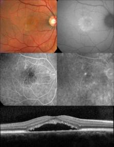
          Advances in optical coherence tomography have enabled a better appreciation of the role of pathologic choroidal changes in a variety of retinal disease. A “pachychoroid” (pachy-[prefix]: thick) is defined as an abnormal and permanent increase in choroidal thickness often showing dilated choroidal vessels and other structural alterations of the normal choroidal architecture. Central serous chorioretinopathy is just one of several pachychoroid-related macular disorders. This review summarizes the current state of knowledge of the pachycoroid spectrum and the hallmark features seen with multimodal imaging analysis of these entities

          Related collections

          Most cited references29

          • Record: found
          • Abstract: found
          • Article: not found

          Optical coherence tomography.

          A technique called optical coherence tomography (OCT) has been developed for noninvasive cross-sectional imaging in biological systems. OCT uses low-coherence interferometry to produce a two-dimensional image of optical scattering from internal tissue microstructures in a way that is analogous to ultrasonic pulse-echo imaging. OCT has longitudinal and lateral spatial resolutions of a few micrometers and can detect reflected signals as small as approximately 10(-10) of the incident optical power. Tomographic imaging is demonstrated in vitro in the peripapillary area of the retina and in the coronary artery, two clinically relevant examples that are representative of transparent and turbid media, respectively.
            Bookmark
            • Record: found
            • Abstract: found
            • Article: not found

            A pilot study of enhanced depth imaging optical coherence tomography of the choroid in normal eyes.

            To measure macular choroidal thickness in normal eyes at different points using enhanced depth imaging (EDI) optical coherence tomography (OCT) and to evaluate the association of choroidal thickness and age. Retrospective, observational case series. EDI OCT images were obtained in patients without significant retinal or choroidal pathologic features. The images were obtained by positioning a spectral-domain OCT device close enough to the eye to acquire an inverted image. Seven sections were obtained within a 5 x 30-degree area centered at the fovea, with 100 scans averaged for each section. The choroid was measured from the outer border of the retinal pigment epithelium to the inner scleral border at 500-microm intervals of a horizontal section from 3 mm temporal to the fovea to 3 mm nasal to the fovea. Statistical analysis was performed to evaluate variations of choroidal thickness at each location and to correlate choroidal thickness and patient age. The mean age of the 30 patients (54 eyes) was 50.4 years (range, 19 to 85 years), and 14 patients (46.7%) were female. The choroid was thickest underneath the fovea (mean, 287 microm; standard deviation, +/- 76 microm). Choroidal thickness decreased rapidly in the nasal direction and averaged 145 microm (+/- 57 microm) at 3 mm nasal to the fovea. Increasing age was correlated significantly with decreasing choroidal thickness at all points measured. Regression analysis suggested that the subfoveal choroidal thickness decreased by 15.6 microm for each decade of life. Choroidal thickness seems to vary topographically within the posterior pole. The thickness of the choroid showed a negative correlation with age. The decrease in the thickness of the choroid may play a role in the pathophysiologic features of various age-related ocular conditions.
              Bookmark
              • Record: found
              • Abstract: found
              • Article: not found

              Proposed lexicon for anatomic landmarks in normal posterior segment spectral-domain optical coherence tomography: the IN•OCT consensus.

              To develop a consensus nomenclature for the classification of retinal and choroidal layers and bands visible on spectral-domain optical coherence tomography (SD-OCT) images of a normal eye.
                Bookmark

                Author and article information

                Journal
                Med Hypothesis Discov Innov Ophthalmol
                mehdiophth
                Medical Hypothesis, Discovery and Innovation in Ophthalmology
                Medical Hypothesis, Discovery & Innovation Ophthalmology
                2322-4436
                2322-3219
                Winter 2014
                : 3
                : 4
                : 111-115
                Affiliations
                [1 ] Unit of Macula, Department of Ophthalmology, University and Polytechnic Hospital La Fe, Valencia, Spain
                [2 ] RETICS Oftared, Health Institute Carlos III, Madrid, Spain
                [3 ] Department of Ophthalmology, University of Santiago de Compostela, Santiago de Compostela, Spain
                [4 ]Ophthalmic Institute Gomez-Ulla, Santiago de Compostela, Spain
                [5 ]Vitreous, Retina, Macula Consultants of New York, New York, USA
                [6 ]LuEsther T. Mertz Retinal Research Center, Manhattan Eye, Ear, and Throat Hospital, New York, USA
                [7 ]Department of Ophthalmology, New York University School of Medicine, New York, USA
                Author notes
                Correspondence to: Dr. Roberto Gallego-Pinazo; Tel: +34659954094; Fax: +34963923470; E-Mail: robertogallego@comv.es
                Article
                mehdiophth-3-111
                4352204
                25756060
                7146069c-9b9b-4dbc-aad3-212ea619924f

                This is an Open Access article distributed under the terms of the Creative Commons Attribution License, ( http://creativecommons.org/licenses/by/3.0/) which permits unrestricted use, distribution, and reproduction in any medium, provided the original work is properly cited.

                History
                Categories
                Review Article

                pachychoroid diseases,macula,central serous chorioretinopathy

                Comments

                Comment on this article