1
views
0
recommends
+1 Recommend
0 collections
    0
    shares
      • Record: found
      • Abstract: found
      • Article: not found

      Advances in Deep Learning for Tuberculosis Screening using Chest X-rays: The Last 5 Years Review

      review-article

      Read this article at

      ScienceOpenPublisherPMC
      Bookmark
          There is no author summary for this article yet. Authors can add summaries to their articles on ScienceOpen to make them more accessible to a non-specialist audience.

          Abstract

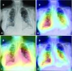
          There has been an explosive growth in research over the last decade exploring machine learning techniques for analyzing chest X-ray (CXR) images for screening cardiopulmonary abnormalities. In particular, we have observed a strong interest in screening for tuberculosis (TB). This interest has coincided with the spectacular advances in deep learning (DL) that is primarily based on convolutional neural networks (CNNs). These advances have resulted in significant research contributions in DL techniques for TB screening using CXR images. We review the research studies published over the last five years (2016-2021). We identify data collections, methodical contributions, and highlight promising methods and challenges. Further, we discuss and compare studies and identify those that offer extension beyond binary decisions for TB, such as region-of-interest localization. In total, we systematically review 54 peer-reviewed research articles and perform meta-analysis.

          Related collections

          Most cited references38

          • Record: found
          • Abstract: found
          • Article: not found

          Deep Learning at Chest Radiography: Automated Classification of Pulmonary Tuberculosis by Using Convolutional Neural Networks

          Purpose To evaluate the efficacy of deep convolutional neural networks (DCNNs) for detecting tuberculosis (TB) on chest radiographs. Materials and Methods Four deidentified HIPAA-compliant datasets were used in this study that were exempted from review by the institutional review board, which consisted of 1007 posteroanterior chest radiographs. The datasets were split into training (68.0%), validation (17.1%), and test (14.9%). Two different DCNNs, AlexNet and GoogLeNet, were used to classify the images as having manifestations of pulmonary TB or as healthy. Both untrained and pretrained networks on ImageNet were used, and augmentation with multiple preprocessing techniques. Ensembles were performed on the best-performing algorithms. For cases where the classifiers were in disagreement, an independent board-certified cardiothoracic radiologist blindly interpreted the images to evaluate a potential radiologist-augmented workflow. Receiver operating characteristic curves and areas under the curve (AUCs) were used to assess model performance by using the DeLong method for statistical comparison of receiver operating characteristic curves. Results The best-performing classifier had an AUC of 0.99, which was an ensemble of the AlexNet and GoogLeNet DCNNs. The AUCs of the pretrained models were greater than that of the untrained models (P < .001). Augmenting the dataset further increased accuracy (P values for AlexNet and GoogLeNet were .03 and .02, respectively). The DCNNs had disagreement in 13 of the 150 test cases, which were blindly reviewed by a cardiothoracic radiologist, who correctly interpreted all 13 cases (100%). This radiologist-augmented approach resulted in a sensitivity of 97.3% and specificity 100%. Conclusion Deep learning with DCNNs can accurately classify TB at chest radiography with an AUC of 0.99. A radiologist-augmented approach for cases where there was disagreement among the classifiers further improved accuracy. © RSNA, 2017.
            Bookmark
            • Record: found
            • Abstract: found
            • Article: found
            Is Open Access

            Iteratively Pruned Deep Learning Ensembles for COVID-19 Detection in Chest X-Rays

            We demonstrate use of iteratively pruned deep learning model ensembles for detecting pulmonary manifestations of COVID-19 with chest X-rays. This disease is caused by the novel Severe Acute Respiratory Syndrome Coronavirus 2 (SARS-CoV-2) virus, also known as the novel Coronavirus (2019-nCoV). A custom convolutional neural network and a selection of ImageNet pretrained models are trained and evaluated at patient-level on publicly available CXR collections to learn modality-specific feature representations. The learned knowledge is transferred and fine-tuned to improve performance and generalization in the related task of classifying CXRs as normal, showing bacterial pneumonia, or COVID-19-viral abnormalities. The best performing models are iteratively pruned to reduce complexity and improve memory efficiency. The predictions of the best-performing pruned models are combined through different ensemble strategies to improve classification performance. Empirical evaluations demonstrate that the weighted average of the best-performing pruned models significantly improves performance resulting in an accuracy of 99.01% and area under the curve of 0.9972 in detecting COVID-19 findings on CXRs. The combined use of modality-specific knowledge transfer, iterative model pruning, and ensemble learning resulted in improved predictions. We expect that this model can be quickly adopted for COVID-19 screening using chest radiographs.
              Bookmark
              • Record: found
              • Abstract: found
              • Article: not found

              Truncated inception net: COVID-19 outbreak screening using chest X-rays

              Since December 2019, the Coronavirus Disease (COVID-19) pandemic has caused world-wide turmoil in a short period of time, and the infection, caused by SARS-CoV-2, is spreading rapidly. AI-driven tools are used to identify Coronavirus outbreaks as well as forecast their nature of spread, where imaging techniques are widely used, such as CT scans and chest X-rays (CXRs). In this paper, motivated by the fact that X-ray imaging systems are more prevalent and cheaper than CT scan systems, a deep learning-based Convolutional Neural Network (CNN) model, which we call Truncated Inception Net, is proposed to screen COVID-19 positive CXRs from other non-COVID and/or healthy cases. To validate our proposal, six different types of datasets were employed by taking the following CXRs: COVID-19 positive, Pneumonia positive, Tuberculosis positive, and healthy cases into account. The proposed model achieved an accuracy of 99.96% (AUC of 1.0) in classifying COVID-19 positive cases from combined Pneumonia and healthy cases. Similarly, it achieved an accuracy of 99.92% (AUC of 0.99) in classifying COVID-19 positive cases from combined Pneumonia, Tuberculosis, and healthy CXRs. To the best of our knowledge, as of now, the achieved results outperform the existing AI-driven tools for screening COVID-19 using the acquired CXRs, and proves the viability of using the proposed Truncated Inception Net as a screening tool.
                Bookmark

                Author and article information

                Contributors
                santosh.kc@usd.edu
                sivasaivenkata.allu@coyotes.usd.edu
                sivaramakrishnan.rajaraman@nih.gov
                sameer.antani@nih.gov
                Journal
                J Med Syst
                J Med Syst
                Journal of Medical Systems
                Springer US (New York )
                0148-5598
                1573-689X
                15 October 2022
                2022
                : 46
                : 11
                : 82
                Affiliations
                [1 ]GRID grid.267169.d, ISNI 0000 0001 2293 1795, Applied Artificial Intelligence (2AI) Research Lab Computer Science Department, , University of South Dakota, ; Vermillion, SD 57069 USA
                [2 ]GRID grid.94365.3d, ISNI 0000 0001 2297 5165, National Library of Medicine, , National Institutes of Health, ; Bethesda, MD 20894 USA
                Author information
                http://orcid.org/0000-0003-4176-0236
                http://orcid.org/0000-0003-0871-8634
                http://orcid.org/0000-0002-0040-1387
                Article
                1870
                10.1007/s10916-022-01870-8
                9568934
                36241922
                1d555011-d288-494b-ae13-4267d9c955b1
                © The Author(s), under exclusive licence to Springer Science+Business Media, LLC, part of Springer Nature 2022, Springer Nature or its licensor holds exclusive rights to this article under a publishing agreement with the author(s) or other rightsholder(s); author self-archiving of the accepted manuscript version of this article is solely governed by the terms of such publishing agreement and applicable law.

                This article is made available via the PMC Open Access Subset for unrestricted research re-use and secondary analysis in any form or by any means with acknowledgement of the original source. These permissions are granted for the duration of the World Health Organization (WHO) declaration of COVID-19 as a global pandemic.

                History
                : 20 January 2022
                : 19 September 2022
                Categories
                Image & Signal Processing
                Custom metadata
                © Springer Science+Business Media, LLC, part of Springer Nature 2022

                Public health
                tuberculosis,chest x-rays,deep learning,medical imaging,systematic review
                Public health
                tuberculosis, chest x-rays, deep learning, medical imaging, systematic review

                Comments

                Comment on this article