11
views
0
recommends
+1 Recommend
0 collections
    0
    shares
      • Record: found
      • Abstract: found
      • Article: found
      Is Open Access

      Collagen V-induced nasal tolerance downregulates pulmonary collagen mRNA gene and TGF-beta expression in experimental systemic sclerosis

      research-article

      Read this article at

      Bookmark
          There is no author summary for this article yet. Authors can add summaries to their articles on ScienceOpen to make them more accessible to a non-specialist audience.

          Abstract

          Background

          The purpose of this study was to evaluate collagen deposition, mRNA collagen synthesis and TGF-beta expression in the lung tissue in an experimental model of scleroderma after collagen V-induced nasal tolerance.

          Methods

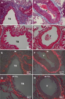
          Female New Zealand rabbits (N = 12) were immunized with 1 mg/ml of collagen V in Freund's adjuvant (IM). After 150 days, six immunized animals were tolerated by nasal administration of collagen V (25 μg/day) (IM-TOL) daily for 60 days. The collagen content was determined by morphometry, and mRNA expressions of types I, III and V collagen were determined by Real-time PCR. The TGF-beta expression was evaluated by immunostaining and quantified by point counting methods. To statistic analysis ANOVA with Bonferroni test were employed for multiple comparison when appropriate and the level of significance was determined to be p < 0.05.

          Results

          IM-TOL, when compared to IM, showed significant reduction in total collagen content around the vessels (0.371 ± 0.118 vs. 0.874 ± 0.282, p < 0.001), bronchioles (0.294 ± 0.139 vs. 0.646 ± 0.172, p < 0.001) and in the septal interstitium (0.027 ± 0.014 vs. 0.067 ± 0.039, p = 0.026). The lung tissue of IM-TOL, when compared to IM, showed decreased immunostaining of types I, III and V collagen, reduced mRNA expression of types I (0.10 ± 0.07 vs. 1.0 ± 0.528, p = 0.002) and V (1.12 ± 0.42 vs. 4.74 ± 2.25, p = 0.009) collagen, in addition to decreased TGF-beta expression (p < 0.0001).

          Conclusions

          Collagen V-induced nasal tolerance in the experimental model of SSc regulated the pulmonary remodeling process, inhibiting collagen deposition and collagen I and V mRNA synthesis. Additionally, it decreased TGF-beta expression, suggesting a promising therapeutic option for scleroderma treatment.

          Related collections

          Most cited references26

          • Record: found
          • Abstract: found
          • Article: not found

          Oral tolerance: immune mechanisms and the generation of Th3-type TGF-beta-secreting regulatory cells.

          Oral tolerance is a long recognized method to induce peripheral immune tolerance. Oral tolerance has been used successfully to treat animal models of autoimmune diseases and is being tested in human diseases. Low doses of oral antigen induce active suppression, whereas high doses induce clonal anergy and deletion. Oral antigen preferentially generates a Th2(IL-4/IL-10)- or a Th3(TGF-beta)-type response. Th3-type cells are a unique T-cell subset which primarily secrete TGF-beta, provide help for IgA and have suppressive properties for Th1 and other immune cells. Th3-type cells appear distinct from the Th2 cells as CD4(+) TGF-beta-secreting cells with suppressive properties in the gut have been generated from IL-4-deficient animals. In vitro differentiation of Th3-type cells from Th0 precursors from TCR transgenic mice is enhanced by culture with TGF-beta, IL-4, IL-10 and anti-IL-12. Because regulatory T cells generated by oral antigen are triggered in an antigen-specific fashion but suppress in an antigen-nonspecific fashion, they mediate bystander suppression when they encounter the fed autoantigen at the target organ. Thus, mucosal tolerance can be used to treat inflammatory processes that are not autoimmune in nature. Mucosal antigen has also been used to treat animal models of stroke and of Alzheimer's disease. Induction of low-dose oral tolerance is enhanced by oral administration of IL-4 and IL-10. Coupling antigen to CTB or administration of Flt-3 ligand enhances oral tolerance. Anti-B7.2 but not anti-B7.1 blocks low-dose, but not high-dose oral tolerance. High-dose oral tolerance is blocked by anti-CTLA-4. CD25(+) CD4(+) regulatory T-cell function also appears to be related to TFG-beta.
            Bookmark
            • Record: found
            • Abstract: found
            • Article: not found

            Oligoclonal T cell expansion in the skin of patients with systemic sclerosis.

            Fibrosis, microvascular fibroproliferative alterations, and autoantibody production are the main features of systemic sclerosis (SSc), and all of them can be explained by cytokine production by activated T cells. However, little is known about the role of T cells in the pathogenesis of SSc, and there is no information on the Ag(s) that elicits such activation. To determine whether T cells infiltrating the skin biopsies of patients with SSc are oligoclonal, beta-chain TCR transcripts from T cells infiltrating the skin of five patients with SSc of recent onset were amplified by either Vbeta-specific PCR or nonpalindromic adaptor PCR. The resulting PCR products were subsequently cloned and sequenced. High proportions of identical beta-chain TCR transcripts ranging from 43 to 90% of those sequenced were found in five patients, strongly suggesting the presence of oligoclonal T cells in these infiltrates. A dominant T cell clone was found to be clonally expanded in skin biopsies obtained from a single patient with SSc at three different times (0, 8, and 13 mo earlier) and from three different skin regions. beta-chain TCR transcripts from PBMC from normal donors (methodological control) were unique when compared with each other, typical for polyclonal populations of T cells. The finding of oligoclonal T cells infiltrating the skin of patients with SSc suggests that these T cells have undergone proliferation in situ in the skin and clonal expansion in response to as yet unidentified Ag(s). These results suggest that T cells are involved in the pathogenesis of the disease.
              Bookmark
              • Record: found
              • Abstract: found
              • Article: not found

              Autocrine TGF-beta signaling in the pathogenesis of systemic sclerosis.

              Excessive extracellular matrix deposition in the skin, lung, and other organs is a hallmark of systemic sclerosis (SSc). Fibroblasts isolated from sclerotic lesions in patients with SSc and cultured in vitro are characterized by increased synthesis of collagen and other extracellular matrix components, consistent with the disease phenotype. Thus, cultured scleroderma fibroblasts serve as a principal experimental model for studying the mechanisms involved in extracellular matrix overproduction in SSc. The pathogenesis of SSc is still poorly understood, but increasing evidence suggests that transforming growth factor-beta (TGF-beta) is a key mediator of tissue fibrosis as a consequence of extracellular matrix accumulation in the pathology of SSc. TGF-beta regulates diverse biological activities including cell growth, cell death or apoptosis, cell differentiation, and extracellular matrix synthesis. TGF-beta is known to induce the expression of extracellular matrix proteins in mesenchymal cells and to stimulate the production of protease inhibitors that prevent enzymatic breakdown of the extracellular matrix. This review focuses on the possible role of autocrine TGF-beta signaling in the pathogenesis of SSc.
                Bookmark

                Author and article information

                Journal
                Respir Res
                Respiratory Research
                BioMed Central
                1465-9921
                1465-993X
                2010
                4 January 2010
                : 11
                : 1
                : 1
                Affiliations
                [1 ]Rheumatology Division of the School of Medicine of the University of São Paulo (FMUSP), São Paulo, SP, Brazil
                [2 ]Department of Radiology, Discipline of Oncology of the School of Medicine of the University of São Paulo (FMUSP), São Paulo, SP, Brazil
                [3 ]Department of Pathology of the School of Medicine of the University of São Paulo (FMUSP), São Paulo, SP, Brazil
                Article
                1465-9921-11-1
                10.1186/1465-9921-11-1
                2817654
                20047687
                1bf3da3d-2d43-4cfd-bf7b-530820d731d8
                Copyright ©2010 Velosa et al; licensee BioMed Central Ltd.

                This is an Open Access article distributed under the terms of the Creative Commons Attribution License ( http://creativecommons.org/licenses/by/2.0), which permits unrestricted use, distribution, and reproduction in any medium, provided the original work is properly cited.

                History
                : 21 September 2009
                : 4 January 2010
                Categories
                Research

                Respiratory medicine
                Respiratory medicine

                Comments

                Comment on this article