16
views
0
recommends
+1 Recommend
0 collections
    0
    shares
      • Record: found
      • Abstract: found
      • Article: found
      Is Open Access

      Type V Collagen Induced Tolerance Suppresses Collagen Deposition, TGF-β and Associated Transcripts in Pulmonary Fibrosis

      research-article

      Read this article at

      Bookmark
          There is no author summary for this article yet. Authors can add summaries to their articles on ScienceOpen to make them more accessible to a non-specialist audience.

          Abstract

          Rationale

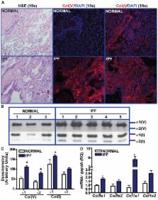
          Idiopathic pulmonary fibrosis (IPF) is a fatal interstitial lung disease characterized by progressive scarring and matrix deposition. Recent reports highlight an autoimmune component in IPF pathogenesis. We have reported anti-col(V) immunity in IPF patients. The objective of our study was to determine the specificity of col(V) expression profile and anti-col(V) immunity relative to col(I) in clinical IPF and the efficacy of nebulized col(V) in pre-clinical IPF models.

          Methods

          Col(V) and col(I) expression profile was analyzed in normal human and IPF tissues. C57-BL6 mice were intratracheally instilled with bleomycin (0.025 U) followed by col(V) nebulization at pre-/post-fibrotic stage and analyzed for systemic and local responses.

          Results

          Compared to normal lungs, IPF lungs had higher protein and transcript expression of the alpha 1 chain of col(V) and col(I). Systemic anti-col(V) antibody concentrations, but not of anti-col(I), were higher in IPF patients. Nebulized col(V), but not col(I), prevented bleomycin-induced fibrosis, collagen deposition, and myofibroblast differentiation. Col(V) treatment suppressed systemic levels of anti-col(V) antibodies, IL-6 and TNF-α; and local Il-17a transcripts. Compared to controls, nebulized col(V)-induced tolerance abrogated antigen-specific proliferation in mediastinal lymphocytes and production of IL-17A, IL-6, TNF-α and IFN-γ. In a clinically relevant established fibrosis model, nebulized col(V) decreased collagen deposition. mRNA array revealed downregulation of genes specific to fibrosis ( Tgf-β, Il-1β, Pdgfb), matrix ( Acta2, Col1a2, Col3a1, Lox, Itgb1/6, Itga2/3) and members of the TGF-β superfamily ( Tgfbr1/2, Smad2/3, Ltbp1, Serpine1, Nfkb/Sp1/Cebpb).

          Conclusions

          Anti-col(V) immunity is pathogenic in IPF, and col(V)-induced tolerance abrogates bleomycin-induced fibrogenesis and down regulates TGF- β-related signaling pathways.

          Related collections

          Most cited references32

          • Record: found
          • Abstract: found
          • Article: not found

          Pulmonary fibrosis: pathogenesis, etiology and regulation

          Pulmonary fibrosis and architectural remodeling of tissues can severely disrupt lung function, often with fatal consequences. The etiology of pulmonary fibrotic diseases is varied, with an array of triggers including allergens, chemicals, radiation and environmental particles. However, the cause of one of the most common pulmonary fibrotic conditions, idiopathic pulmonary fibrosis (IPF), is still unclear. This review examines common mechanisms of pulmonary wound-healing responses following lung injury, and highlights the pathogenesis of some of the most widespread pulmonary fibrotic diseases. A three phase model of wound repair is reviewed that includes; (1) injury; (2) inflammation; and (3) repair. In most pulmonary fibrotic conditions dysregulation at one or more of these phases has been reported. Chronic inflammation can lead to an imbalance in the production of chemokines, cytokines, growth factors, and disrupt cellular recruitment. These changes coupled with excessive pro-fibrotic IL-13 and/or TGFβ1 production can turn a well-controlled healing response into a pathogenic fibrotic response. Endogenous regulatory mechanisms are discussed including novel areas of therapeutic intervention. Restoring homeostasis to these dysregulated healing responses, or simply neutralizing the key pro-fibrotic mediators may prevent or slow the progression of pulmonary fibrosis.
            Bookmark
            • Record: found
            • Abstract: found
            • Article: not found

            NADPH Oxidase-4 Mediates Myofibroblast Activation and Fibrogenic Responses to Lung Injury

            The NADPH oxidase (NOX) family of enzymes, which catalyze the reduction of O2 to form reactive oxygen species (ROS), have increased in number during eukaryotic evolution1,2. Seven isoforms of the NOX gene family have been identified in mammals; however, specific roles of NOX enzymes in mammalian physiology and pathophysiology have not been fully elucidated3,4. The best established physiological role of NOX enzymes is in host defense against pathogen invasion in diverse species, including plants5,6. The prototypical member of this family, NOX2 (gp91 phox ), is expressed in phagocytic cells and mediates microbicidal activities7,8. Here, we report a role for the NOX4 isoform in tissue repair functions of myofibroblasts and fibrogenesis. Transforming growth factor-β1 (TGF-β1) induces NOX4 expression in lung mesenchymal cells by a SMAD3-dependent mechanism. NOX4-dependent generation of hydrogen peroxide (H2O2) is required for TGF-β1-induced myofibroblast differentiation, extracellular matrix (ECM) production, and contractility. NOX4 is upregulated in lungs of mice subjected to non-infectious injury and in human idiopathic pulmonary fibrosis (IPF). Genetic or pharmacologic targeting of NOX4 abrogates fibrogenesis in two different murine models of lung injury. These studies support a novel function for NOX4 in tissue fibrogenesis and provide proof-of-concept for therapeutic targeting of NOX4 in recalcitrant fibrotic disorders.
              Bookmark
              • Record: found
              • Abstract: found
              • Article: not found

              Adenovector-mediated gene transfer of active transforming growth factor-beta1 induces prolonged severe fibrosis in rat lung.

              Transforming growth factor (TGF)-beta1 has been implicated in the pathogenesis of fibrosis based upon its matrix-inducing effects on stromal cells in vitro, and studies demonstrating increased expression of total TGF-beta1 in fibrotic tissues from a variety of organs. The precise role in vivo of this cytokine in both its latent and active forms, however, remains unclear. Using replication-deficient adenovirus vectors to transfer the cDNA of porcine TGF-beta1 to rat lung, we have been able to study the effect of TGF-beta1 protein in the respiratory tract directly. We have demonstrated that transient overexpression of active, but not latent, TGF-beta1 resulted in prolonged and severe interstitial and pleural fibrosis characterized by extensive deposition of the extracellular matrix (ECM) proteins collagen, fibronectin, and elastin, and by emergence of cells with the myofibroblast phenotype. These results illustrate the role of TGF-beta1 and the importance of its activation in the pulmonary fibrotic process, and suggest that targeting active TGF-beta1 and steps involved in TGF-beta1 activation are likely to be valuable antifibrogenic therapeutic strategies. This new and versatile model of pulmonary fibrosis can be used to study such therapies.
                Bookmark

                Author and article information

                Contributors
                Role: Editor
                Journal
                PLoS One
                PLoS ONE
                plos
                plosone
                PLoS ONE
                Public Library of Science (San Francisco, USA )
                1932-6203
                2013
                21 October 2013
                : 8
                : 10
                : e76451
                Affiliations
                [1 ]Center for Immunobiology, Indiana University School of Medicine, Indianapolis, Indiana, United States of America
                [2 ]ImmuneWorks Inc., Indianapolis, Indiana, United States of America
                [3 ]Department of Pathology, Indiana University School of Medicine, Indianapolis, Indiana, United States of America
                Helmholtz Zentrum München/Ludwig-Maximilians-University Munich, Germany
                Author notes

                Competing Interests: David S. Wilkes is a co-founder of ImmuneWorks, Inc., a biotechnology company involved in developing therapeutics for various forms of lung diseases. Katia Rothhaar is the Director of Research Operations while Sarah Frye is a Research Technician working at ImmuneWorks, Inc. All other authors declare no competing interests. This does not alter the authors' adherence to all the PLOS ONE policies on sharing data and materials.

                Conceived and designed the experiments: RV DSW. Analyzed the data: RV KR DSW. Wrote the paper: RV DSW. Pepsin digestion of clinical tissues, hydroxyproline analyses: EAM. Bleomycin instillations: AF. Real-time PCR on patient and animal tissues: CZ HG. Col(V) antibody analyses on patient and murine plasma: KR KMB SF. Patient demographic analyses: AE. Cytokine bead assay: JML. Received IPF tissues from LTRC: RV. Provided consultation on col(V) pepsin digestion: GNS. Board-certified pathologists: OWC GES. Patient demographic analyses: AE. Cytokine bead assay: JML.

                Article
                PONE-D-12-36671
                10.1371/journal.pone.0076451
                3804565
                8fd8e03f-71d9-4307-9003-192463c40a2e
                Copyright @ 2013

                This is an open-access article distributed under the terms of the Creative Commons Attribution License, which permits unrestricted use, distribution, and reproduction in any medium, provided the original author and source are credited.

                History
                : 29 November 2012
                : 29 August 2013
                Page count
                Pages: 14
                Funding
                This work was supported by NIH-NHLBI-Lung Tissue Research Consortium and NIH-NHLBI-HL109288 to RV; NIH-NCAATS-KL2 TR000163 (A. Shekhar, PI; Co-investigator: RV); and by the NIH-NHLBI-HL067177 and NIAID P01AI084853 to DSW. The funders had no role in study design, data collection and analysis, decision to publish, or preparation of the manuscript.
                Categories
                Research Article

                Uncategorized
                Uncategorized

                Comments

                Comment on this article