29
views
0
recommends
+1 Recommend
0 collections
    0
    shares
      • Record: found
      • Abstract: found
      • Article: found
      Is Open Access

      Acid Stress Response Mechanisms of Group B Streptococci

      review-article

      Read this article at

      Bookmark
          There is no author summary for this article yet. Authors can add summaries to their articles on ScienceOpen to make them more accessible to a non-specialist audience.

          Abstract

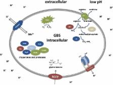
          Group B streptococcus (GBS) is a leading cause of neonatal mortality and morbidity in the United States and Europe. It is part of the vaginal microbiota in up to 30% of pregnant women and can be passed on to the newborn through perinatal transmission. GBS has the ability to survive in multiple different host niches. The pathophysiology of this bacterium reveals an outstanding ability to withstand varying pH fluctuations of the surrounding environments inside the human host. GBS host pathogen interations include colonization of the acidic vaginal mucosa, invasion of the neutral human blood or amniotic fluid, breaching of the blood brain barrier as well as survival within the acidic phagolysosomal compartment of macrophages. However, investigations on GBS responses to acid stress are limited. Technologies, such as whole genome sequencing, genome-wide transcription and proteome mapping facilitate large scale identification of genes and proteins. Mechanisms enabling GBS to cope with acid stress have mainly been studied through these techniques and are summarized in the current review

          Related collections

          Most cited references140

          • Record: found
          • Abstract: found
          • Article: not found

          Genome analysis of multiple pathogenic isolates of Streptococcus agalactiae: implications for the microbial "pan-genome".

          The development of efficient and inexpensive genome sequencing methods has revolutionized the study of human bacterial pathogens and improved vaccine design. Unfortunately, the sequence of a single genome does not reflect how genetic variability drives pathogenesis within a bacterial species and also limits genome-wide screens for vaccine candidates or for antimicrobial targets. We have generated the genomic sequence of six strains representing the five major disease-causing serotypes of Streptococcus agalactiae, the main cause of neonatal infection in humans. Analysis of these genomes and those available in databases showed that the S. agalactiae species can be described by a pan-genome consisting of a core genome shared by all isolates, accounting for approximately 80% of any single genome, plus a dispensable genome consisting of partially shared and strain-specific genes. Mathematical extrapolation of the data suggests that the gene reservoir available for inclusion in the S. agalactiae pan-genome is vast and that unique genes will continue to be identified even after sequencing hundreds of genomes.
            Bookmark
            • Record: found
            • Abstract: found
            • Article: not found

            Fluorescence probe measurement of the intralysosomal pH in living cells and the perturbation of pH by various agents.

            A quantitative method is described for the measurement of intralysosomal pH in living cells. Fluorescein isothiocyanate-labeled dextran (FD) is endocytized and accumulates in lysosomes where it remains without apparent degradation. The fluorescence spectrum of this compound changes with pH in the range 4-7 and is not seriously affected by FD concentration, ionic strength, or protein concentration. Living cells on coverslips are mounted in a spectrofluorometer cell and can be perfused with various media. The normal pH inside macrophage lysosomes seems to be 4.7-4.8, although it can drop transiently as low as 4.5. Exposure of the cells to various weak bases and to acidic potassium ionophores causes the pH to increase. The changes in pH are much more rapid than is the intralysosomal accumulation of the weak bases. Inhibitors of glycolysis (2-deoxyglucose) and of oxidative phosphorylation (cyanide or azide) added together, but not separately, cause the intralysosomal pH to increase. These results provide evidence for the existence of an active proton accumulation mechanism in the lysosomal membrane and support the theory of lysosomal accumulation of weak bases by proton trapping.
              Bookmark
              • Record: found
              • Abstract: found
              • Article: not found

              Uptake and synthesis of compatible solutes as microbial stress responses to high-osmolality environments.

              All microorganisms possess a positive turgor, and maintenance of this outward-directed pressure is essential since it is generally considered as the driving force for cell expansion. Exposure of microorganisms to high-osmolality environments triggers rapid fluxes of cell water along the osmotic gradient out of the cell, thus causing a reduction in turgor and dehydration of the cytoplasm. To counteract the outflow of water, microorganisms increase their intracellular solute pool by amassing large amounts of organic osmolytes, the so-called compatible solutes. These osmoprotectants are highly congruous with the physiology of the cell and comprise a limited number of substances including the disaccharide trehalose, the amino acid proline, and the trimethylammonium compound glycine betaine. The intracellular amassing of compatible solutes as an adaptive strategy to high-osmolality environments is evolutionarily well-conserved in Bacteria, Archaea, and Eukarya. Furthermore, the nature of the osmolytes that are accumulated during water stress is maintained across the kingdoms, reflecting fundamental constraints on the kind of solutes that are compatible with macromolecular and cellular functions. Generally, compatible solutes can be amassed by microorganisms through uptake and synthesis. Here we summarise the molecular mechanisms of compatible solute accumulation in Escherichia coli and Bacillus subtilis, model organisms for the gram-negative and gram-positive branches of bacteria.
                Bookmark

                Author and article information

                Contributors
                Journal
                Front Cell Infect Microbiol
                Front Cell Infect Microbiol
                Front. Cell. Infect. Microbiol.
                Frontiers in Cellular and Infection Microbiology
                Frontiers Media S.A.
                2235-2988
                07 September 2017
                2017
                : 7
                : 395
                Affiliations
                [1] 1Institute of Medical Microbiology and Hygiene, University of Ulm Ulm, Germany
                [2] 2Department of Microbiology and Immunology, Faculty of Pharmacy, Suez Canal University Ismailia, Egypt
                Author notes

                Edited by: Wolfgang Eisenreich, Technische Universität München, Germany

                Reviewed by: Peter Valentin-Weigand, University of Veterinary Medicine Hannover, Germany; Sven Hammerschmidt, University of Greifswald, Germany

                *Correspondence: Barbara Spellerberg barbara.spellerberg@ 123456uniklinik-ulm.de
                Article
                10.3389/fcimb.2017.00395
                5594096
                28936424
                1b22278e-4af4-4575-be62-4a50440ded8a
                Copyright © 2017 Shabayek and Spellerberg.

                This is an open-access article distributed under the terms of the Creative Commons Attribution License (CC BY). The use, distribution or reproduction in other forums is permitted, provided the original author(s) or licensor are credited and that the original publication in this journal is cited, in accordance with accepted academic practice. No use, distribution or reproduction is permitted which does not comply with these terms.

                History
                : 26 March 2017
                : 23 August 2017
                Page count
                Figures: 2, Tables: 0, Equations: 0, References: 154, Pages: 13, Words: 12161
                Funding
                Funded by: Ministry of Higher Education 10.13039/501100002385
                Categories
                Microbiology
                Review

                Infectious disease & Microbiology
                streptococcus agalactiae,acid resistance,low ph,molecular mechanisms,stress response

                Comments

                Comment on this article