11
views
0
recommends
+1 Recommend
1 collections
    0
    shares

      International Journal of Nanomedicine (submit here)

      This international, peer-reviewed Open Access journal by Dove Medical Press focuses on the application of nanotechnology in diagnostics, therapeutics, and drug delivery systems throughout the biomedical field. Sign up for email alerts here.

      105,621 Monthly downloads/views I 7.033 Impact Factor I 10.9 CiteScore I 1.22 Source Normalized Impact per Paper (SNIP) I 1.032 Scimago Journal & Country Rank (SJR)

      • Record: found
      • Abstract: found
      • Article: found
      Is Open Access

      Rapid Detection Method for Pathogenic Candida Captured by Magnetic Nanoparticles and Identified Using SERS via AgNPs +

      research-article

      Read this article at

      ScienceOpenPublisherPMC
      Bookmark
          There is no author summary for this article yet. Authors can add summaries to their articles on ScienceOpen to make them more accessible to a non-specialist audience.

          Abstract

          Purpose

          Candidemia infection is common in the clinic and has a high mortality rate. Candida albicans, Candida tropicalis, and Candida krusei are very important and common pathogenic species. Candida is difficult to isolate from clinical samples and culture, and immunological detection cannot distinguish these related strains. Furthermore, Candida has a complex cell wall, which causes difficulties in the extraction of DNA for nucleic acid detection. The purpose of this study was to establish a protocol for the direct identification of Candida from serum.

          Materials and Methods

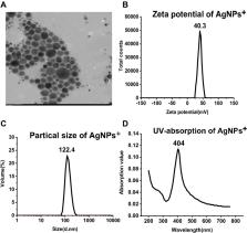
          We synthesized Fe 3O 4@PEI (where PEI stands for polyethylenimine) magnetic nanoparticles to capture Candida and prepared positively charged silver nanoparticles (AgNPs +) as the substrate for surface-enhanced Raman scattering (SERS). Candida was directly identified from serum by SERS detection.

          Results

          Orthogonal partial least squares discriminant analysis (OPLS-DA) was used as the multivariate analysis tool. Principal component analysis confirmed that this method can clearly distinguish common Candida. After 10-fold cross-validation, the accuracy of training data in this model was 100% and the accuracy of test data was 99.8%, indicating that the model has good classification ability.

          Conclusion

          The detection could be completed within 40 minutes using Fe 3O 4@PEI and AgNPs + prepared in advance. This is the first time that Fe 3O 4@PEI was used in the detection of Candida by SERS. We report the first rapid method to identify fungi directly from serum without breaking the cell wall to extract DNA from the fungi.

          Most cited references31

          • Record: found
          • Abstract: found
          • Article: found
          Is Open Access

          Re-epithelialization and immune cell behaviour in an ex vivo human skin model

          A large body of literature is available on wound healing in humans. Nonetheless, a standardized ex vivo wound model without disruption of the dermal compartment has not been put forward with compelling justification. Here, we present a novel wound model based on application of negative pressure and its effects for epidermal regeneration and immune cell behaviour. Importantly, the basement membrane remained intact after blister roof removal and keratinocytes were absent in the wounded area. Upon six days of culture, the wound was covered with one to three-cell thick K14+Ki67+ keratinocyte layers, indicating that proliferation and migration were involved in wound closure. After eight to twelve days, a multi-layered epidermis was formed expressing epidermal differentiation markers (K10, filaggrin, DSG-1, CDSN). Investigations about immune cell-specific manners revealed more T cells in the blister roof epidermis compared to normal epidermis. We identified several cell populations in blister roof epidermis and suction blister fluid that are absent in normal epidermis which correlated with their decrease in the dermis, indicating a dermal efflux upon negative pressure. Together, our model recapitulates the main features of epithelial wound regeneration, and can be applied for testing wound healing therapies and investigating underlying mechanisms.
            Bookmark
            • Record: found
            • Abstract: found
            • Article: not found

            Microwave absorption enhancement of multifunctional composite microspheres with spinel Fe3 O4 Cores and Anatase TiO2 shells.

            Multifunctional composite microspheres with spinel Fe(3)O(4) cores and anatase TiO(2) shells (Fe(3)O(4)@TiO(2)) are synthesized by combining a solvothermal reaction and calcination process. The size, morphology, microstructure, phase purity, and magnetic properties are characterized by scanning electron microscopy, transmission electron microscopy (TEM), high-resolution TEM, selected-area electron diffraction, electron energy loss spectroscopy, powder X-ray diffraction, and superconducting quantum interference device magnetometry. The results show that the as-synthesized microspheres have a unique morphology, uniform size, good crystallinity, favorable superparamagnetism, and high magnetization. By varying the experimental conditions such as Fe(3)O(4) size and concentration, microspheres with different core sizes and shell thickneses can be readily synthesized. Furthermore, the microwave absorption properties of these microspheres are investigated in terms of complex permittivity and permeability. By integration of the chemical composition and unique structure, the Fe(3)O(4)@TiO(2) microspheres possess lower reflection loss and a wider absorption frequency range than pure Fe(3)O(4). Moreover, the electromagnetic data demonstrate that Fe(3)O(4@TiO(2) microspheres with thicker TiO(2) shells exhibit significantly enhanced microwave absorption properties compared to those with thinner TiO(2) shells, which may result from effective complementarities between dielectric loss and magnetic loss. All the results indicate that these Fe(3)O(4)@TiO(2) microspheres may be attractive candidate materials for microwave absorption applications. Copyright © 2012 WILEY-VCH Verlag GmbH & Co. KGaA, Weinheim.
              Bookmark
              • Record: found
              • Abstract: found
              • Article: not found

              Understanding the Promises and Hurdles of Metagenomic Next-Generation Sequencing as a Diagnostic Tool for Infectious Diseases

              Metagenomic next-generation sequencing (mNGS) has emerged as a promising single, universal pathogen detection method for infectious disease diagnostics. With the development of mNGS assays, it is essential for treating practitioners to understand both the power and limitations of the method as a diagnostic tool.
                Bookmark

                Author and article information

                Journal
                Int J Nanomedicine
                Int J Nanomedicine
                ijn
                intjnano
                International Journal of Nanomedicine
                Dove
                1176-9114
                1178-2013
                11 February 2021
                2021
                : 16
                : 941-950
                Affiliations
                [1 ]Department of Biotechnology, Beijing Institute of Radiation Medicine, Beijing Key Laboratory of New Molecular Diagnosis Technologies for Infectious Diseases , Beijing, 100850, People’s Republic of China
                [2 ]Department of Laboratory Medicine, Xuzhou Tumor Hospital , Xuzhou, 221005, People’s Republic of China
                [3 ]Xuzhou Key Laboratory of Laboratory Diagnostics, Medical Technology School of Xuzhou Medical University , Xuzhou, 221004, People’s Republic of China
                [4 ]Department of Laboratory Medicine, The Affiliated Hospital of Xuzhou Medical University , Xuzhou, 221006, People’s Republic of China
                [5 ]College of Life Sciences, Anhui Agricultural University , Hefei, 230036, People’s Republic of China
                Author notes
                Correspondence: Ying Yang; Bing Gu Email y_ying_77@163.com; gb20031129@163.com
                [*]

                These authors contributed equally to this work

                Author information
                http://orcid.org/0000-0002-7096-6527
                Article
                285339
                10.2147/IJN.S285339
                7884937
                14dfa3ca-a366-46b3-8b57-014c08912dd1
                © 2021 Hu et al.

                This work is published and licensed by Dove Medical Press Limited. The full terms of this license are available at https://www.dovepress.com/terms.php and incorporate the Creative Commons Attribution – Non Commercial (unported, v3.0) License ( http://creativecommons.org/licenses/by-nc/3.0/). By accessing the work you hereby accept the Terms. Non-commercial uses of the work are permitted without any further permission from Dove Medical Press Limited, provided the work is properly attributed. For permission for commercial use of this work, please see paragraphs 4.2 and 5 of our Terms ( https://www.dovepress.com/terms.php).

                History
                : 05 October 2020
                : 19 January 2021
                Page count
                Figures: 6, Tables: 3, References: 31, Pages: 10
                Categories
                Original Research

                Molecular medicine
                capture,surface-enhanced raman scattering,positively charged silver nanoparticles,orthogonal partial least squares discriminant analysis,10-fold cross-validation

                Comments

                Comment on this article