35
views
0
recommends
+1 Recommend
0 collections
    0
    shares
      • Record: found
      • Abstract: found
      • Article: found
      Is Open Access

      The Therapeuatic Effect of Endostar on Soft Carotid Plaque Neovascularization in Patients with Non-small Cell Lung Cancer

      research-article

      Read this article at

      Bookmark
          There is no author summary for this article yet. Authors can add summaries to their articles on ScienceOpen to make them more accessible to a non-specialist audience.

          Abstract

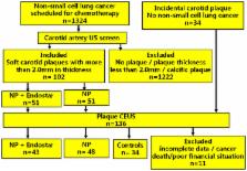
          The purpose of this study was to investigate the effect of the angiogenesis inhibitor Endostar on carotid plaque neovascularization in patients with non-small cell lung cancer (NSCLC) using contrast-enhanced ultrasound (CEUS). Ninety-one patients who had NSCLC with soft carotid plaques were selected for treatment either with the NP regimen (vinorelbine + cisplatin) (43 patients) or with the ENP regimen (Endostar + NP) (48 patients). Plaque thickness and neovascularization of the plaque were assessed before and at 1 month after treatment using CEUS. Enhanced intensity (EI) of CEUS was used for quantification of plaque neovascularization. There was no significant changes in any group in thickness of plaque between recruitment and 1 month after treatment ( P > 0.05 for all). There was no significant change in the EI of plaque in the controls or NP groups at 1 month after treatment ( P > 0.05), while EI in the ENP group was significantly reduced at 1 month after treatment ( P < 0.01) and significantly lower than that in the controls or NP group at 1 month after treatment ( P < 0.001 both). This study indicates that carotid soft plaque neovascularization in patients with NSCLC can be reduced by anti-angiogenesis treatment.

          Related collections

          Most cited references25

          • Record: found
          • Abstract: found
          • Article: not found

          Plaque neovascularization is increased in ruptured atherosclerotic lesions of human aorta: implications for plaque vulnerability.

          Growth of atherosclerotic plaques is accompanied by neovascularization from vasa vasorum microvessels extending through the tunica media into the base of the plaque and by lumen-derived microvessels through the fibrous cap. Microvessels are associated with plaque hemorrhage and may play a role in plaque rupture. Accordingly, we tested this hypothesis by investigating whether microvessels in the tunica media, the base of the plaque, and the fibrous cap are increased in ruptured atherosclerotic plaques in human aorta. Microvessels, defined as CD34-positive tubuloluminal capillaries recognized in cross-sectional and longitudinal profiles, were quantified in 269 advanced human plaques by bicolor immunohistochemistry. Macrophages/T lymphocytes and smooth muscle cells were defined as CD68/CD3-positive and alpha-actin-positive cells. Total microvessel density was increased in ruptured plaques when compared with nonruptured plaques (P=0.0001). Furthermore, microvessel density was increased in lesions with severe macrophage infiltration at the fibrous cap (P=0.0001) and at the shoulders of the plaque (P=0.0001). In addition, microvessel density was also increased in lesions with intraplaque hemorrhage (P=0.04) and in thin-cap fibroatheromas (P=0.038). Logistic regression analysis identified plaque base microvessel density (P=0.003) as an independent correlate to plaque rupture. Thus, neovascularization as manifested by the localized appearance of microvessels is increased in ruptured plaques in the human aorta. Furthermore, microvessel density is increased in lesions with inflammation, with intraplaque hemorrhage, and in thin-cap fibroatheromas. Microvessels at the base of the plaque are independently correlated with plaque rupture, suggesting a contributory role for neovascularization in the process of plaque rupture.
            Bookmark
            • Record: found
            • Abstract: found
            • Article: not found

            Angiogenesis inhibitors endostatin or TNP-470 reduce intimal neovascularization and plaque growth in apolipoprotein E-deficient mice.

            Neovascularization within the intima of human atherosclerotic lesions is well described, but its role in the progression of atherosclerosis is unknown. In this report, we first demonstrate that intimal vessels occur in advanced lesions of apolipoprotein E-deficient (apoE -/-) mice. To test the hypothesis that intimal vessels promote atherosclerosis, we investigated the effect of angiogenesis inhibitors on plaque growth in apoE -/- mice. ApoE -/- mice were fed a 0.15% cholesterol diet. At age 20 weeks, mice were divided into 3 groups and treated for 16 weeks as follows: group 1, recombinant mouse endostatin, 20 mg. kg-1. d-1; group 2, fumagillin analogue TNP-470, 30 mg/kg every other day; and group 3, control animals that received a similar volume of buffer. Average cholesterol levels were similar in all groups. Plaque areas were quantified at the aortic origin. Median plaque area before treatment was 0.250 mm2 (range, 0.170 to 0.348; n=10). Median plaque areas were 0.321 (0.238 to 0.412; n=10), 0.402 (0.248 to 0.533; n=15), and 0.751 mm2 (0.503 to 0.838; n=12) for the endostatin, TNP-470, and control groups, respectively (P
              Bookmark
              • Record: found
              • Abstract: found
              • Article: not found

              Blood pressure rise following angiogenesis inhibition by bevacizumab. A crucial role for microcirculation.

              Arterial hypertension (HT) has been reported in all studies involving bevacizumab, an antiangiogenic agent designed to target vascular endothelial growth factor (VEGF). The mechanism underlying bevacizumab-related HT is not yet clearly understood. As far as endothelial dysfunction and microvascular rarefaction are hallmarks in all forms of HT, we tested the hypothesis that anti-VEGF therapy could alter the microcirculation in nontumor tissues and, thus, result in an increase in blood pressure (BP). We used intravital video microscopy to measure dermal capillary densities in the dorsum of the fingers. Microvascular endothelial function was assessed by laser Doppler flowmetry combined with iontophoresis of pilocarpine (acetylcholine analogue). All measurements were carried out in 18 patients before and after a 6-month treatment with bevacizumab (mean cumulative dose: 3.16 +/- 0.90 g). Mean BP was increased after 6 months of therapy compared with baseline, from 129 +/- 13/75 +/- 7 mmHg to 145 +/- 17/82 +/- 7 mmHg for systolic BP and diastolic BP, respectively (P < 0.0001). Compared with the baseline, mean dermal capillary density at 6 months was significantly lower (75 +/- 12 versus 83 +/- 13/mm(2); P < 0.0001), as well as pilocarpine-induced vasodilation (P < 0.05). Thus, bevacizumab treatment resulted in endothelial dysfunction and capillary rarefaction; both changes are closely associated and could be responsible for the rise in BP observed in most patients.
                Bookmark

                Author and article information

                Journal
                Sci Rep
                Sci Rep
                Scientific Reports
                Nature Publishing Group
                2045-2322
                10 March 2015
                2015
                : 5
                : 8956
                Affiliations
                [1 ]Department of Ultrasound, the Second Affiliated Hospital of Zhejiang University School of Medicine , Zhejiang 310009, China
                [2 ]Department of Ultrasound, the Second Affiliated Hospital of Wenzhou Medical University , Zhejiang 325027, China
                [3 ]Baylor Heart & Vascular Institute, Baylor University Medical Center , 621 N. Hall St., Suite H030 Dallas, Texas 75226, USA
                Author notes
                Article
                srep08956
                10.1038/srep08956
                4354169
                25753083
                145df9c2-2cf4-4c63-9398-b06bb23ecd4e
                Copyright © 2015, Macmillan Publishers Limited. All rights reserved

                This work is licensed under a Creative Commons Attribution 4.0 International License. The images or other third party material in this article are included in the article's Creative Commons license, unless indicated otherwise in the credit line; if the material is not included under the Creative Commons license, users will need to obtain permission from the license holder in order to reproduce the material. To view a copy of this license, visit http://creativecommons.org/licenses/by/4.0/

                History
                : 02 December 2014
                : 12 February 2015
                Categories
                Article

                Uncategorized
                Uncategorized

                Comments

                Comment on this article