2
views
0
recommends
+1 Recommend
0 collections
    0
    shares
      • Record: found
      • Abstract: found
      • Article: found
      Is Open Access

      Conidiobolomycose (entomophthoromycose rhinofaciale) au Gabon, à propos d'une observation Translated title: Conidiobolomycosis (rhinofacial entomophthoromycosis) in Gabon. About of one case

      research-article

      Read this article at

      Bookmark
          There is no author summary for this article yet. Authors can add summaries to their articles on ScienceOpen to make them more accessible to a non-specialist audience.

          Résumé

          Introduction

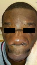
          La conidiobolomycose ou entomophthoromycose rhinofaciale est une mycose sous-cutanée tropicale rare, réalisant dans les formes évoluées un aspect dysmorphique, typique, du visage en « museau d'hippopotame », dont peu de cas ont été rapportés dans la littérature.

          Méthodologie

          Nous présentons l'observation d'un patient de 25 ans, vivant en zone équatoriale, au sud du Gabon en environnement forestier humide.

          Résultats

          Les données histologiques de la biopsie cutanée associées à la présentation clinique étaient compatibles avec le diagnostic de conidiobolomycose. L’évolution initiale était favorable sur le plan esthétique sous itraconazole 300 mg/jour pendant 2 mois et corticothérapie (bolus de méthylprednisone 240 mg/jour pendant 3 jours relayée per os à la dose de 0,5 mg/kg/jour (soit 30 mg/jour) de prednisone), maintenue pendant 3 mois. L'amélioration moyenne nasale n'a pu être complétée par une chirurgie et le malade a été perdu de vue.

          Conclusion

          Cette deuxième observation de conidiobolomycose au Gabon dans la même province, fait de la Ngounié, un écosystème privilégié de cette affection.

          Translated abstract

          Background

          Entomophthoromycosis constitutes a nosological group of subcutaneous mycoses including conidiobolomycosis (rhinofacial form) and basidiobomomycosis (subcutaneous form involving the trunk and the limbs). Conidiobolomycosis is characterized by a progressive nasal and facial deformity giving, in the evolved forms, a “hippopotamus snout”. The literature review finds a hundred cases, with a tropism for the humid tropical regions. Methods. We report the observation of a 25-year-old patient, living in the equatorial zone, in the south of Gabon in a humid forest area, presenting a swollen aspect of the face mainly involving the eyelids, the nose and the upper lips.

          Results

          The diagnosis of entomophthoromycosis was compatible with the histopathological and clinical aspects. The evolution was favorable in terms of facial aesthetics under itraconazole 300 mg/day for 2 months and corticosteroid therapy (bolus of methylprednisone 240 mg/day for 3 days relayed per os at a dose of 0.5 mg/kg/day, i.e. 30 mg/day) of prednisone), maintained for 3 months. The average nasal improvement could not be completed by surgery and the patient was lost to follow-up.

          Conclusion

          This second observation of conidiobolomycosis in Gabon in the same province makes Ngounié a privileged ecosystem for this affection.

          Related collections

          Most cited references14

          • Record: found
          • Abstract: found
          • Article: not found

          Human Pathogenic Entomophthorales

          SUMMARY The pathogenic entomophthoralean fungi cause infection in insects and mammalian hosts. Basidiobolus and Conidiobolus species can be found in soil and insect, reptile, and amphibian droppings in tropical and subtropical areas. The life cycles of these fungi occur in these environments where infecting sticky conidia are developed. The infection is acquired by insect bite or contact with contaminated environments through open skin. Conidiobolus coronatus typically causes chronic rhinofacial disease in immunocompetent hosts, whereas some Conidiobolus species can be found in immunocompromised patients. Basidiobolus ranarum infection is restricted to subcutaneous tissues but may be involved in intestinal and disseminated infections. Its early diagnosis remains challenging due to clinical similarities to other intestinal diseases. Infected tissues characteristically display eosinophilic granulomas with the Splendore-Höeppli phenomenon. However, in immunocompromised patients, the above-mentioned inflammatory reaction is absent. Laboratory diagnosis includes wet mount, culture serological assays, and molecular methodologies. The management of entomophthoralean fungi relies on traditional antifungal therapies, such as potassium iodide (KI), amphotericin B, itraconazole, and ketoconazole, and surgery. These species are intrinsically resistant to some antifungals, prompting physicians to experiment with combinations of therapies. Research is needed to investigate the immunology of entomophthoralean fungi in infected hosts. The absence of an animal model and lack of funding severely limit research on these fungi.
            Bookmark
            • Record: found
            • Abstract: found
            • Article: not found

            Rhinofacial conidiobolomycosis (entomophthoramycosis).

            Conidiobolomycosis is a tropical rhinofacial subcutaneous mycosis caused by Conidiobolus coronatus. It is characterized histopathologically by the presence of hyphae surrounded by an eosinophilic halo, the so-called Splendore-Hoeppli phenomenon. There is no standard treatment, but itraconazole is an effective option.
              Bookmark
              • Record: found
              • Abstract: found
              • Article: not found

              Conidiobolomycosis in a young Malaysian woman showing chronic localized fibrosing leukocytoclastic vasculitis: a case report and meta-analysis focusing on clinicopathologic and therapeutic correlations with outcome.

              Conidiobolomycosis (also known as rhinoentomophthoramycosis) is a rare cutaneous/mucosal fungal infection seen mainly in the tropical rain forest regions of the world that can be associated with disfiguring facial elephantiasis, and rarely, death.
                Bookmark

                Author and article information

                Contributors
                Journal
                Med Trop Sante Int
                Med Trop Sante Int
                MTSI
                Médecine Tropicale et Santé Internationale
                MTSI
                2778-2034
                2778-2034
                31 December 2023
                13 December 2023
                : 3
                : 4
                : mtsi.v3i4.2023.457
                Affiliations
                [1 ]Service de dermatologie, CHU de Libreville, Libreville, Gabon
                [2 ]Service de médecine interne, CHU de Libreville, Libreville, Gabon
                Author notes
                Article
                10.48327/mtsi.v3i4.2023.457
                10879889
                38390015
                0efeac9a-bd09-4ee2-b9f3-602dad915a46
                Copyright © 2023 SFMTSI

                Cet article en libre accès est distribué selon les termes de la licence Creative Commons CC BY 4.0 ( https://creativecommons.org/licenses/by/4.0/).

                History
                : 12 May 2023
                : 28 November 2023
                Page count
                Figures: 4, References: 14, Pages: 5
                Categories
                Cas Clinique
                Clinical Case

                conidiobolomycose,entomophthoromycose rhinofaciale,itraconazole,amélioration clinique,la ngounié,gabon,afrique subsaharienne,conidiobolomycosis,rhinofacial entomophtoromycosis,clinical improvement,ngounié province,sub-saharan africa

                Comments

                Comment on this article